Download
1 / 23
230 likes | 740 Vues
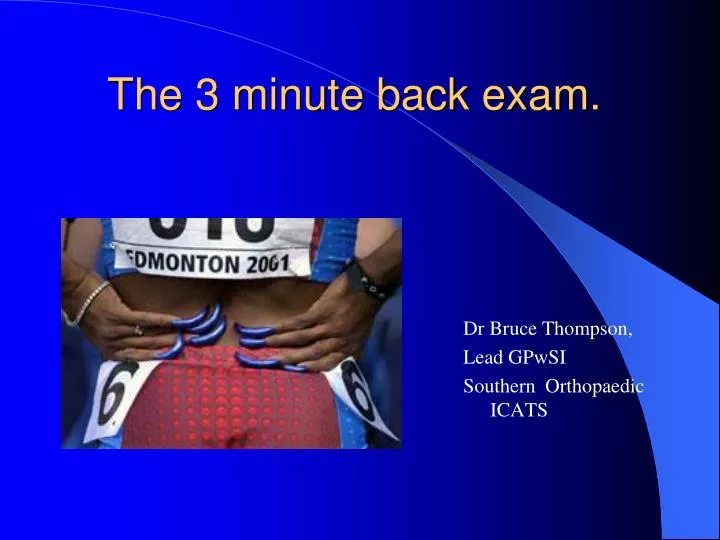
The 3 minute back exam. Dr Bruce Thompson, Lead GPwSI Southern Orthopaedic ICATS. “Back pain”. Very vague non-specific term “Abdominal pain” would not be described in general terms for diagnosis or treatment.

E N D
The 3 minute back exam. Dr Bruce Thompson, Lead GPwSI Southern Orthopaedic ICATS
“Back pain” • Very vague non-specific term • “Abdominal pain” would not be described in general terms for diagnosis or treatment. • Need to be as specific as possible – consider inflammatory, spasmodic, neuropathic, mechanical etc • Evidence basis suffers for being non-specific – unsuitable treatments applied to patients
Sources of back pain • Intervertebral disc – outer 1/3rd • Vertebra – body or posterior structures • Muscles • Thoracolumbar fascia • Dura mater • Epidural plexus • Ligaments • Joints – facet or sacroiliac • Intra-abdominal
Discogenic pain • Internal disc disruption is the cardinal pathological basis for lumbar discogenic pain. • The prevalence of IDD is 40% in patients with chronic LBP • Diagnosis is by +ve disc stimulation at affected level with –ve stimulation above/below
Disc facts • If disc narrowed more force goes on facets • 75% of disc hydration lost in 1st hour of waking – protect spine then as 18% loss of strength • Compressive force endplate # • Facets limit torsion but can get rim tear of disc • Degeneration often genetic – FH of OA • Discs are largest avascular structures in the body
Dural referred pain • Dural stimuli cause referred pain to be felt in the “pantaloon” distribution.
Gross classification of LBP • Red flag problem • Radiculopathy – acute or chronic • Mechanical – acute or chronic
Red Flags (a) • Age <20 or > 55 years • Violent trauma ? # • Constant progressive non-mechanical pain • Thoracic pain • PMH Carcinoma • Systemic steroids • Drug abuse / HIV
Red Flags (b) • Systemically unwell, weight loss • Saddle anaesthesia, GIT / GUS upset • Persisting severe limitation of flexion • Widespread neurological deficit • Structural deformity
History - ? emotional language • Age and occupation – exact details • Sports and training / coaching – exact details • Onset and duration • Site and spread • Symptoms – factors affecting • Other joint problems • PMH / FH • Drugs • Treatment to date – who, what, when.
General Observation • Face • Posture • Gait • Simple movements • Activities
Inspection • Bony deformity • Colour changes • Wasting • Swelling • Scars
Active Movements • - for pain, range and willingness – in lumbar spine active “flows” into passive due to gravity • Extension • Side flexion • Forward flexion
Tests while standing • Single leg standing – Trendelenburg’s sign • Calf raise – unilateral • One-legged hyperextension - spondylolysis • Flexibility – quadriceps, hamstrings, adductors, calf muscles
Supine • Hip range of movement • Sacro-iliac joint tests – FABER & shear • Straight leg raising – add bias • Sensation • Reflexes
Lumbosacral dermatomes • L4 – big toe • L5 – lateral toes • S1 – stand on • S2 – slip on • S3 – sit on • S4 - perianal
Myotomes – maximum force to test power and pain • L2 Psoas - hip flexion • L3 Quadriceps - knee extension • L4 Tibialis anterior - ankle dorsiflexion • L5 Ext. Hall. Long. - big toe dorsiflexion • S1 Peroneals - ankle eversion
Prone • Hip extension Femoral stretch test • Sacral compression • Lumbar extension – McKenzie Test • Lumbar vertebral extension thrusts • Gluteal muscle tone
Dynamic tests • Quick test- drop to hunkering then rise • Muscle endurance of flexors, extensors, side flexors • Gun-dog, side bridge, bridging • Core stability balance and co-ordination • Shear stability test
Malingering? • Juddering movements • Flip test – SLR then sit with legs extended • Hoover test – cup calcaneus in each hand and feel counter-pressure on SLR • Axial loading on head / cervical spine • Simulated rotation – of legs not spine
Beware! • The bizarre but persistent patient