Download
1 / 20
200 likes | 732 Vues
Serguey A. Khoruzhik, MD Computed Tomography , Grodno Regional Clinical Hospital , BLK 52, 2300 17 Grodno, Belarus Tel.: + (375 152) 331320 khoruzhik@grsmi.unibel.by http://nld.hut.ru/e/cv.htm. The 60th Jubilee Congress of The Association of Polish Surgeons, Warsaw 12-15.IX.2001.

E N D
SergueyA. Khoruzhik, MDComputed Tomography, Grodno Regional Clinical Hospital,BLK 52, 230017 Grodno, BelarusTel.: + (375 152) 331320khoruzhik@grsmi.unibel.byhttp://nld.hut.ru/e/cv.htm The 60th Jubilee Congress of The Association of Polish Surgeons, Warsaw 12-15.IX.2001 SYSTEMIC HEMANGIOMATOSIS WITH ATYPICAL LIVER HEMANGIOMAS AND DIAPHRAGM INVOLVEMENT Khoruzhik S. A.1, Maslakova N. D.2, Siljaeva N. F.3From The 1Departments of Radiology, 2Abdominal Surgery, and 3Pathology, Grodno Regional Clinical Hospital, Grodno, Belarus Contact:
Grodno Regional Clinical Hospital, Grodno, Belarus INTRODUCTION • Haemangioma is the most common benign hepatic tumor, being present in about 5 % of livers autopsy. • In systemic haemangiomatosis multiple organs involved including liver, spleen, muscles, bowel, lungs, brain. • On unenhanced CT haemangioma usually appears as well-marginated solid mass of the same or decreased relative to liver parenchyma density what corresponds to hyperechoic lesion on US.
Grodno Regional Clinical Hospital, Grodno, Belarus INTRODUCTION CT scan without contrast enhancement shows typical haemangioma in the 4th liver segment.
Grodno Regional Clinical Hospital, Grodno, Belarus CASE REPORT: history • A 68-year-old female presented with abdominal pain, nausea, weakness, intermittent temperature of 38° C and chilling. She was underwent spleenectomy one month earlier in other hospital because of spontaneous spleen rupture followed by subfebrile temperature up to the time of present admission.
Grodno Regional Clinical Hospital, Grodno, Belarus CASE REPORT: imaging findings Post-contrast liver CT scan shows high contrast enhancement in the capsules of multiple nodular lesions measuring 2 to 5 cm in diameter. Central parts of the lesions remain unenhanced.
Grodno Regional Clinical Hospital, Grodno, Belarus CASE REPORT: imaging findings Post-contrast liver CT scan. Free fluid in the both spleen bed and left pleural cavity present. Left hemidiaphragm appeared irregularly thickened.
Grodno Regional Clinical Hospital, Grodno, Belarus CASE REPORT: imaging findings This corresponded to hyperechoic ring-shaped structures with anechoic centre on US exam.
Grodno Regional Clinical Hospital, Grodno, Belarus CASE REPORT: diagnosis? • Multiple liver abscesses Based on imaging findings and history of resent operation on abdomen first diagnostic choice was: Second choice was: • Cystic metastases
Grodno Regional Clinical Hospital, Grodno, Belarus CASE REPORT: diagnosis? • Hemorrhagic fluid was aspirated from the liver lesion under CT guidance what raised suspicion of haemangiomas. • 1,5 litres of hemorrhagic fluid was aspirated from the left pleural cavity. Explorative thoracotomy revealed diffuse bleeding from the left hemidiaphragm into pleural cavity. • Spleen microphotographs were revised and proliferation of vascular channels characteristic for haemangiomas was found.
Grodno Regional Clinical Hospital, Grodno, Belarus CASE REPORT: outcome • During next few weeks patient’s condition deteriorated rapidly with clinic of sepsis and disseminated intravascular coagulation. In one month after admission to the hospital patient died. • On post-mortem study multiple cavernous haemangiomas were found in liver, epiploon, abdominal ligaments, and left diaphragmatic muscle. All liver lesions contained large central areas of intratumoral hemorrhage.
Grodno Regional Clinical Hospital, Grodno, Belarus CASE REPORT: final diagnosis Systemic haemangiomatosis with multiple liver cavernous haemangiomas and left diaphragmatic musclehaemangiomas with clinical presentation of Kasabach-Merrit syndrome.
Grodno Regional Clinical Hospital, Grodno, Belarus DIFFERENTIAL DIAGNOSIS OF LIVER CYSTIC MASSES • Abscesses. • Cystic metastases. • Atypical liver haemangiomas. • Hepatocellular carcinoma with cystic change. • Hepatic tumor after treatment (embolisation, radio-frequency ablation). • Polycystic liver disease.
Grodno Regional Clinical Hospital, Grodno, Belarus DIFFERENTIAL DIAGNOSIS: atypical liver haemangioma • On US: solid tumor with an echogenic rim and hypoechoic internal echo pattern. • On CT: ring-like structure with hypodense unenhancing centre. • The reasons for central hypoechoity/hypodensity are internal hemorrhage with necrosis, thrombosis, scaring, and myxomatous change. • In one study this kind of lesions were present in 15 of 5000 abdominal US exams (0.3 % prevalence of atypical haemangiomas in the general population).
Grodno Regional Clinical Hospital, Grodno, Belarus DIFFERENTIAL DIAGNOSIS: abscess CT scans of the liver in two different patients. Abscess in the right liver lobe (left) and atypical liver haemangiomas (right) for comparison.
Grodno Regional Clinical Hospital, Grodno, Belarus DIFFERENTIAL DIAGNOSIS: abscess Liver CT scan shows hepatic abscesses in the right lobe with blood products precipitated along the posterior wall. In such instance blood might be aspirated from the abscess.
Grodno Regional Clinical Hospital, Grodno, Belarus DIFFERENTIAL DIAGNOSIS: cystic metastases Most often liver metastases are solid. Cystic metastases are uncommon and may arise from mucin-producing primaries or be due to necrosis within the tumor in: • ovarian carcinoma, • colorectal carcinoma, • melanoma, • cervical carcinoma, • leiomyosarcoma, • lung cancer.
Grodno Regional Clinical Hospital, Grodno, Belarus DIFFERENTIAL DIAGNOSIS: cystic metastases CT scan shows multiple metastases from colorectal carcinoma. Some of the lesions demonstrates central hypodensity.
Grodno Regional Clinical Hospital, Grodno, Belarus DIFFERENTIAL DIAGNOSIS: polycystic liver disease CT scan shows multiple liver cysts. Some of the cysts have calcified walls as consequence of chronic inflammation.
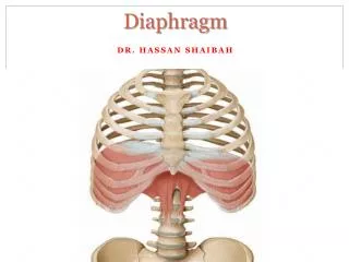
Grodno Regional Clinical Hospital, Grodno, Belarus DISTINCTIVE FIATURES OF THE PRESENTED CASE • Atypical liver haemangiomas simulating liver abscesses or cystic metastases both radiologically and clinically. • Presence of diaphragmatic haemangiomas. Intramuscular haemangiomas are rare benign tumors, making up 0.8% of all haemangiomas. There were just two reports of diaphragmatic haemangiomas in English medical literature.
Grodno Regional Clinical Hospital, Grodno, Belarus CONCLUSION Atypical hepatic cavernous haemangiomas may present on imaging as multiple ring-like non-specific lesions mimicking abscesses and metastases. This diagnosis should be considered in patients with cystic hepatic lesions without primary malignancy. Possibility of diaphragm involvement in systemic haemangiomatosis has to be taken into account.