Download
1 / 14
210 likes | 1.07k Vues
Electrical Impedance and Colorimetric Measurements. Joanna Ellis, MLS(ASCP). Objectives. Cite the electrical impedance principle of cell counting. Identify and interpret microcytic and macrocytic RBC histograms Define coincidence

E N D
Electrical ImpedanceandColorimetric Measurements Joanna Ellis, MLS(ASCP)
Objectives • Cite the electrical impedance principle of cell counting. • Identify and interpret microcytic and macrocytic RBC histograms • Define coincidence • Identify the cell populations represented on a 3-part differential WBC histogram
HistoryYou CAN patent a hole • Prior to the 1950s blood cell counts were performed by manual methods: • Hemacytometer blood counts • Spun hematocrits • Spectrophotometrically determined hemoglobins • Peripheral blood cell evaluation for all differentials • In 1953, Wallace Coulter patented the Coulter Principle in which particles are counted in fluid passed through a hole. The incredulous attorneys who had told him “You can’t patent a hole” were proven wrong. • Hematology automation has since grown to include additional principles such as optical light scatter and flow cytometry.
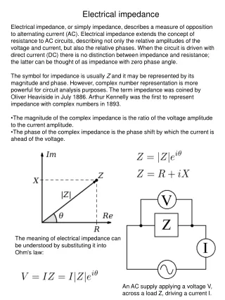
The Coulter Principle • The poorly conductive blood cells are suspended in a conductive diluent (liquid). • The diluent is passed through an electric field created between two electrodes. • The liquid passes through a small aperture (hole). • The passage of each particle through the aperture momentarily increases the impedance (resistance) of the electrical path between the electrodes. • The increase in impedance creates a pulse that can be measured. • The number of pulses = blood cell count • The amplitude (height) of the pulse = Volume of cell
Electrical Impedance (The Coulter Principle) Sweep Flow: Steady stream of diluent that flows behind each aperture to prevent cells from re-entering the aperture
Counting Chambers • Most common chambers using impedance: • RBC/Platelet chamber • WBC chamber RBC/Platelet Chamber WBC Chamber Differential Chamber Reticulocyte Channel
RBC and Platelet Histograms The black line represents normal cell distribution. The red line on the RBC histogram graphically represents a microcytic red cell population.
Bimodal Histogram • Bimodal peak can be seen in situations such • Cold agglutinin disease • Post-transfusion • Post-treatment of IDA
Coincidence Coincidence: Multiple cells passed through the aperture at once.
WBC Chamber WBCs Lysing agent Aperture in electric field Hgb released In some analyzers the WBC count is directly measured by electric impedance after the red cells have been destroyed by a lysing agent. The lysing agent also shrinks the leukocyte cell membrane and cytoplasm; therefore, the WBC count represents the measure of the cell volume not native cell size. Colorimetric measurements are used to determine hemoglobin.
Colorimetric Measurements • Hemoglobin is often determined by a colorimetric method. • Imidazole • Non-cyanide reagent with color change and read at 540nm • Instruments • Abbott CELL-DYN Sapphire • Sodium Lauryl Sulfate • Non-cyanide reagent with color change and read spectrophotometrically • Instruments • Sysmex XT and XE • Lysing agent converts free hemoglobin to cyanmethemoglobin and read spectrophotometrically at 540nm. • Instruments • Advia 120 • Some Beckman Coulters
Three-part Differential WBC Histogram Lymphocytes: 35-92L Mononuclear Cells: 92-152 fL Granulocytes: 152-450 fL
References • "Abaxis Veterinary Diagnostics - VetScan HM2 Technology." Abaxis Medical Diagnostics - Redirect. Abaxis Medical Diagnostics, 2006. Web. 12 Sept. 2010. <http://www.abaxis.com/veterinary/vetscan_hm2_technology.html>. • Graham, Marshall Don. "The Coulter Principle: Foundation of an Industry." The Association for Laboratory Automation :: Home. JALA Volume 8, Issue 6, Dec. 2003. Web. 12 Sept. 2010. <http://labautomation.org>. • Kelly, Kathleen. "Modules :: CLIA :: CME." Hematology in the Physician Office Laboratory Section I. University of Iowa Carver College of Medicine, 2008. Web. 12 Sept. 2010. <http://www.medicine.uiowa.edu/cme/clia/modules.asp?testID=4#02>. • Krantz, Alexander. "Residency & Fellowship Programs | Education." Department of Pathology & Cell Biology | CUMC. Columbia University of Physicians and Surgeons. Web. 12 Sept. 2010. <http://pathology.columbia.edu/education/residency>. • Harmening., Denise, Clinical Hematology and Fundamentals of Hemostasis, 3rd edition, pp. 593-599. • Turgeon, Mary Louise, Clinical Hematology - Theories and Procedures, 3rd edition, pp373, 376-382. • Rodak, Bernadette, Diagnostic Hematology, 1st edition, p.605-606. • Coulter STKS Operating Manual • McKenzie, Shirlyn, Clinical Laboratory Hematology, 2ndedition,pp 813-829.