Download
1 / 51
710 likes | 4.25k Vues
Necrotizing Fasciitis. Elizabeth Ann Feely, M.D. Department of Internal Medicine Resident Grand Rounds December 1, 1998. Case Presentation. HPI : B.T. is a 48 YOWF with ESRD, DM, HTN, and CAD, who was recently hospitalized for uncomplicated

E N D
Necrotizing Fasciitis Elizabeth Ann Feely, M.D. Department of Internal Medicine Resident Grand Rounds December 1, 1998
Case Presentation HPI: B.T. is a 48 YOWF with ESRD, DM, HTN, and CAD, who was recently hospitalized for uncomplicated angioplasty of her LAD. She presented to WFU/BMC ED with c/o a “painful boil on her bottom ” x 3 days. Pain started in the region of her right groin catheterization site and had progressively become unbearable. Mild subjective fever and chills. Denied N/V, SOB, CP, diarrhea, dysuria, or vaginal discharge. Meds: ASA, Atenolol, Imdur, Insulin NPH, Prilosec, Nu-Iron, Folate, and NTG SL
Case Presentation Pertinent Physical Exam Findings: VS: T 96.6, P 56, BP 86/42, R 20 Gen: obese WF in NAD CV/Lungs: within normal limits Abd: soft, obese, slightly tender in right lower quadrant, nl. BS. Large hematoma in right groin w/ surrounding erythema. No purulence. On deep palpation, + soft tissue crepitus. GU: grossly swollen labium, with crepitus on palpation.
10.3 134 89 35 324 28.3 383 9.3 19 3.3 S B L 94 2 2 Case Presentation Labs:
Case Presentation ED Course: Pt. started on KVO NS and given 1 g IV Rocephin. Medicine consulted for ? infected Bartholin’s cyst. Plain film of pelvis was recommended. Plain film showed air within soft tissues from groin into labium. Given her crepitance, WBC, and DM, necrotizing fasciitis was high on differential. Surgery consult performed incision over groin hematoma, revealing foul-smelling, nonviable SQ tissue.
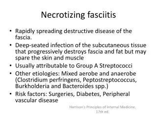
Definition Necrotizing fasciitis (NF) • rare, life-threatening soft-tissue infection usually caused by toxin-producing bacteria • characterized by widespread fascial necrosis with sparing of skin and muscle • can be associated with severe systemic toxicity • may rapidly lead to death unless promptly treated • part of a spectrum of soft-tissue infections, which can be categorized anatomically
Anatomic and Clinical Classification of Soft Tissue Infections
History • Earliest report dates back to 5th century B.C. when Hippocrates described a complication of erysipelas: “… the erysipelas would quickly spread widely in all directions. Flesh, sinews and bones fell away in large quantities… Fever was sometimes present and sometimes absent… There were many deaths. The course of the disease was the same to whatever part of the body it spread.” • first described in U.S. in 1871 • the term, necrotizing fasciitis, first used in 1952
Epidemiology • estimates of 500-1,500 new cases are reported in the US each year ; rare • actual incidence is complicated by use of multiple terms to describe same entity • acute non-clostridial crepitant cellulitis, non-clostridial gas gangrene, synergistic necrotizing cellulitis, necrotizing cellulitis, bacterial synergistic gangrene, gangrenous or necrotizing erysipelas, hemolytic streptococcal gangrene, Fournier’s gangrene. • no age or sex predilection
Bacteriology • ~ 10% of cases due to aerobes, 20% due to anaerobes, 70% due to mixed flora • A single organism is isolated in < 10% of cases. • Two types: Type 1: mixed anaerobic, aerobic, and facultative bacteria; accounts for ~ 90% of cases. Type 2: Group A Streptococcus only; occurs ~ 10% of cases.
Bacteriology • Overall, strep is most common causative organism. • Most cases caused by GAS are M-type 1, 3, 12, or 28. The M protein and exotoxins impede phagocytosis and liberate cytokines, causing a toxic shock-like syndrome.
Risk Factors • Diabetes mellitus • Peripheral vascular disease • IV drug use • Alcoholism • Immunosupressed patients • Old age/obesity/malnutrition
Etiology/Initiating Events • Can affect any part of body • 80% caused by bacteria that extend from contaminated disruptions in skin or localized skin infection • General: skin disruption (cut, abrasion, etc.), blunt/penetrating trauma, post-op complications, cutaneous infections/ulcers, illicit/SQ drug injections • Abdominal Wall: post-op complication of abd surgery
Etiology/Initiating Events • Extremity: trauma, illicit drug use, insect bite, scratch, or wound • Perineum: post-op complication, pilonidal abscess, neglected ischiorectal/perineal abscess • Vulva: Bartholin’s gland duct abscess, vulvar abscess, pudendal nerve block; post-op wound infection from C-section, episiotomy, hysterectomy, etc. • Fournier’s gangrene (NF of male genital organs): GU infections, traumatic instrumentation, urethral calculus, neoplasm, surgery, coital injury
Etiology/Initiating Events • Head and Neck • Scalp/Periorbital: trauma, eyelid infection/puritis • Face/Neck: progressive dental infections, peritonsillar abscess, salivary gland infections, cervical adenitis, otologic sources • Complication of percutaneous catheter placement: chest tube, PEG, and percutaneous drain of abdominal abscess
Etiology/Initiating Events • Idiopathic necrotizing fasciitis • cases that occur without an obvious portal of entry • occur in ~ 20% of cases • more likely to occur in healthy patients • typically caused by single organism (Strep pyogenes) • commonly involves lower extremities • result of infection from unrecognized breaks in skin or hematogenous spread
Clinical Presentation • Initially present with nonspecific symptoms (pain, high fever, edema, and erythema). • Pain out of proportion to exam and systemic toxicity should suggest possibility of NF. • Initial signs include erythematous, swollen, tender area of cellulitis- distinct margins and induration are absent.
Clinical Presentation • With progression, skin color darkens, blisters develop, and skin can become anesthetized with focal areas of skin necrosis. • Fascial necrosis occurs with involved fascia and SQ fat appearing dull grey, edematous, and necrotic with serosanguinous exudate.
Pathophysiology • precise mechanism is not known • thought to be due to bacterial enzymes (lipases and hyaluronidase) • extent of fascial necrosis is more widespread than overlying skin changes • arteries can thrombose, resulting in focal areas of necrosis; nerves can be destroyed • signs of sepsis occur as organisms and toxins are delivered into bloodstream
The Importance of Rapid Diagnosis Study by Lille et al. suggests a delay in the diagnosis by a matter of hours significantly increases mortality. • 10 yr. retrospective analysis of 29 pts. w/ confirmed NF (by intraop findings) • pts. categorized into early group (surgery <24 hours; n=17) or delayed group (surgery >24 hours; n=12) • no significant differences between groups based on age, gender, infection location, or premorbid medical conditions
Importance of Rapid Diagnosis Lille et al. • Mortality was 6% in the early group verses 25% in the delayed group. • Advanced clinical signs were present in 76% of early group verses 25% of the delayed group. • Other factors (FNA, radiographs,etc.) were also compared between the two groups.
Importance of Rapid Diagnosis Lille et al. • Study suggests prompt diagnosis followed by appropriate treatment decreases mortality in pts. w/ NF. • Factors contributing to diagnostic delay were: • lack of clinical symptoms and signs • negative FNA and radiographic changes • admission to a nonsurgical service
Clinical Question What is the best diagnostic modality available to establish the diagnosis of necrotizing fasciitis?
Review of the Literature • Review was performed on the following diagnostic modalities: • frozen tissue biopsy • plain film • computed tomography • magnetic resonance imaging • Limitations were small study sizes. Difficult to perform prospective RCTs because of the rarity of the disease and the benefits to any procedure that could possibly shorten the diagnostic delay.
Frozen Tissue Biopsy Stamenkovic and Lew studied frozen -section biopsy for diagnosis of NF in early stages. • 13 yr. retrospective review of 19 cases of histologically proven NF • Pts. were categorized into frozen-section biopsy group or group who did not undergo biopsy, but had diagnosis made later on clinical grounds or surgical resection. • Differences were seen in the mortality and the time between onset of sxs and surgery.
Frozen Tissue Biopsy Comparative Mortality and Time (Between Onset of Sympotms and Treatment) for Biopsy Group and Non-Biopsied Group • Table 2 mort
Frozen Tissue Biopsy Stamenkovic and Lew • performing frozen-section soft-tissue biopsy early can provide definitive diagnosis and decrease mortality • biopsies excluded the diagnosis in 6 cases • drawbacks
Frozen tissue biopsy Majeski and Majeski performed a similar study. • 43 pts. with similar variety of clinical sxs were evaluated for possible NF with frozen section biopsy. • 12/43 had frozen section diagnosis of NF • 20/43 had cellulitis • 11/43 had abscess w/o advancing necrotizing infection
Frozen Tissue Biopsy Majeski • 100% of pts. W/ NF survived • avg. time between initial eval. and surgery was 4 hours • frozen section biopsy provides reliable diagnosis of NF • drawbacks
Plain Film Fisher et al. studied roentgenographic studies in diagnosing NF. • 9 yr. retrospective review of 26 cases of NF (proven by clinical, gross, and pathologic criteria) • gas was found in soft tissues of 19 pts. (73%) and was seen as bubbles at surgery • crepitation and plain films were checked on each pt. to detect the presence of soft-tissue gas
Plain film • Table 3
Plain film Fisher et al. • physical exam finding of crepitus not a reliable tool to detect soft-tissue gas • x-rays detected gas reliably and simply • drawbacks • x-rays are specific but infrequently show soft-tissue gas
Computed Tomography Wysoki et al. studied computed tomography (CT) characteristics of NF. • 4 yr. retrospective review of 20 preop CT scans of pts. w/ NF (pathologically proven) • CT findings were correlated w/ clinical factors • asymmetric fascial thickening & fat stranding (80%) • gas tracking along fascial planes (55%) • focal fluid collections (35%)
Computed Tomography • CT scan pic
Computed Tomography Wysoki et al. • asymmetric fascial thickening and gas on CT are valuable in diagnosing NF • CT can also delineate true extent of disease • pts. w/o soft-tissue gas on CT create more of a diagnostic challenge • drawbacks
Magnetic Resonance Imaging Schmid et al. evaluated MRI in differentiating NF from cellulitis. • 4 yr. retrospective review of MRI exams on 17 pts. w/ clinically suspected NF • cellulitis diagnosed by thickening and contrast enhancement of SQ tissues/ superficial fascia and deeper structures nl. • NF diagnosed by deep fascial signal intensities (on T2-weighted and contrast enhanced T1-weighted images)
Magnetic Resonance Imaging Schmid et al. • final diagnosis was NF in 11/17 pts. (proven surgically or autopsy) and cellulitis in 6/17 pts. • 11 cases of NF and 5/6 cases of cellulitis were correctly identified • one case of cellulitis was overstaged • sensitivity of 100%, specificity of 86%
Magnetic Resonance Imaging Loh et al. studied specificity of deep fascial hyperintense T2-weighted signal abnormalities for necrotizing soft-tissue infections. • 22 MRIs (w/ deep fascial signal abnl.) retrospectively reviewed w/o knowledge of clinical or pathologic diagnosis • necrotizing soft-tissue process was diagnosed if deep fascial signal abnl. were present
Loh et al. Pretty tables 1 and 2
Magnetic Resonance Imaging Loh et al. • differentiation of NF from other abnormal soft-tissue conditions based on presence of hyperintense T2-weighted signal in deep fascia is unreliable • other disorders have increased water content of soft-tissue and should be distinguished by history and clinical findings