Download
1 / 49
510 likes | 934 Vues
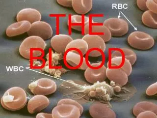
The Blood . Fluids of the Body . Cells of the body utilize 2 fluids Blood composed of plasma and a variety of cells transports nutrients and wastes Interstitial fluid bathes the cells of the body Nutrients and oxygen diffuse from the blood into the interstitial fluid & then into the cells

E N D
Fluids of the Body • Cells of the body utilize 2 fluids • Blood • composed of plasma and a variety of cells • transports nutrients and wastes • Interstitial fluid • bathes the cells of the body • Nutrients and oxygen diffuse from the blood into the interstitial fluid & then into the cells • Wastes move in the reverse direction
Functions of Blood • Transportation • O2, CO2, metabolic wastes, nutrients, heat & hormones • Regulation • helps regulate pH through buffers • helps regulate body temperature • Protection from disease & loss of blood
Physical Characteristics of Blood • Thicker (more viscous) than water and flows more slowly than water • Temperature of 100.40 F • pH 7.4 (7.35-7.45) • Blood volume • 5 to 6 liters in average male • 4 to 5 liters in average female • hormonal negative feedback systems maintain constant blood volume and pressure
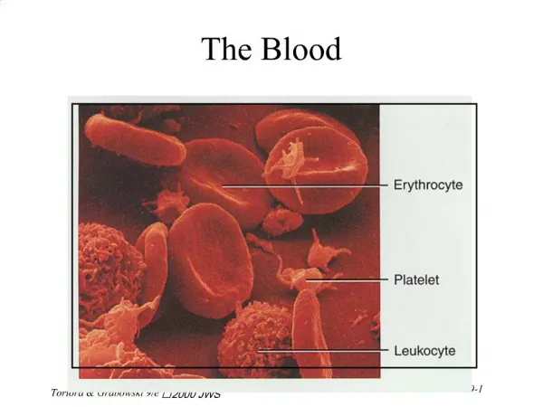
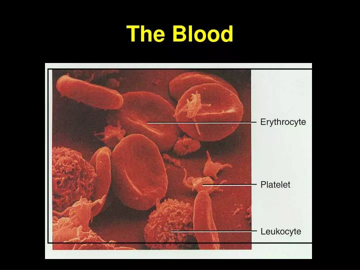
Components of Blood • 55% plasma • 45% cells • 99% RBCs • < 1% WBCs and platelets
Hematocrit • Percentage of blood volume occupied by RBCs • female normal range 38 - 46% (average of 42%) • male normal range 40 - 50% (average of 45%) • Anemia - not enough RBCs • Polycythemia - too many RBCs (over 50%) • Normal hemoglobin range • adult females have 12 to 16 g/100mL of blood • adult males have 13.5 to 18g/100mL of blood
Blood Plasma • Over 90% water • 7% plasma proteins • created in liver • confined to bloodstream • albumin • blood osmotic pressure • transport • globulins (immunoglobulins) • Defense against foreign proteins • fibrinogen • clotting • 2% other substances • electrolytes, nutrients, hormones, gases, waste products
Formed Elements of Blood • Red blood cells ( erythrocytes ) • White blood cells ( leukocytes ) • granular leukocytes • neutrophils • eosinophils • basophils • agranular leukocytes • lymphocytes = T cells, B cells, and natural killer cells • monocytes • One drop of blood: • Normal RBC count ~ 5 million/drop • male 5.4 million/drop • female 4.8 million/drop • WBC count - 5-10,000 white blood cells • Platelet count 150,000-400,000
Formation of Blood Cells • Most blood cell types need to be continually replaced • die within hours, days or weeks • process of blood cells formation is hematopoiesis or hemopoiesis • In adult • occurs only in red marrow of flat bones like sternum, ribs, skull & pelvis and ends of long bones
Red Blood Cells or Erythrocytes • Contain oxygen-carrying protein hemoglobin that gives blood its red color • 1/3 of cell’s weight is hemoglobin • Biconcave disk • increased surface area/volume ratio • flexible shape for narrow passages • no nucleus or other organelles • no mitochondrial ATP formation • New RBCs enter circulation at 2-3 million/second
Hemoglobin • Globin protein consisting of 4 polypeptide chains • One heme pigment attached to each polypeptide chain • each heme contains an iron ion (Fe+2) that can combine reversibly with one oxygen molecule
Function of Hemoglobin • Each hemoglobin molecule can carry 4 oxygen molecules. • Hemoglobin also acts as a buffer and balances pH of blood • Hemoglobin transports 23% of total CO2 waste from tissue cells to lungs for release • combines with amino acids in globin portion of Hb
RBC Life Cycle • RBCs live only 120 days • wear out from bending to fit through capillaries • no repair possible due to lack of organelles • Worn out cells removed by macrophages in spleen & liver • globin portion broken down into amino acids & recycled • heme portion split into iron (Fe+3) and biliverdin (green pigment)
Erythropoiesis: Production of RBCs • Proerythroblast starts to produce hemoglobin • Eventually nucleus is ejected & a reticulocyte is formed • Reticulocytes escape from bone marrow into the blood • In 1-2 days, they eject the remaining organelles to become a mature RBC • Factors needed are erythropoietin from kidneys, Vitamin B12 and Iron
Feedback Control of RBC Production • Tissue hypoxia (cells not getting enough O2) • high altitude since air has less O2 • anemia • RBC production falls below RBC destruction • Kidney response to hypoxia • release erythropoietin • speeds up development of proerythroblasts into reticulocytes
WBC Physiology • Less numerous than RBCs • 5000 to 10,000 cells per drop of blood • 1 WBC for every 700 RBC • Only 2% of total WBC population is in circulating blood at any given time • rest is in lymphatic fluid, skin, lungs, lymph nodes & spleen • Requires colony stimulating factor (local bone marrow/WBC hormone)
Neutrophil Function • Fastest response of all WBC to bacteria and parasites • Direct actions against bacteria • release lysozymes which destroy/digest bacteria • release defensin proteins that act like antibiotics • release strong oxidants (bleach-like, strong chemicals ) that destroy bacteria
Basophil Function • Involved in inflammatory and allergy reactions • Leave capillaries & enter connective tissue as mast cells • Release heparin, histamine & serotonin • heighten the inflammatory response and account for hypersensitivity (allergic) reaction • Heparin is a potent anti-coagulant that does not allow clotting within vessels
Eosinophil Function • Leave capillaries to enter tissue fluid • Release histaminase • slows down inflammation caused by basophils • Attack parasitic worms • Phagocytize antibody-antigen complexes
Monocyte Function • Take longer to get to site of infection, but arrive in larger numbers • Become wandering macrophages, once they leave the capillaries • Destroy microbes and clean up dead tissue following an infection
Lymphocyte Functions • B cells • destroy bacteria and their toxins • turn into plasma cells that produces antibodies • T cells • attack viruses, fungi, transplanted organs, cancer cells • Natural killer cells • attack many different microbes & some tumor cells • destroy foreign invaders by direct attack
Differential WBC Count (FYI) • Detection of changes in numbers of circulating WBCs (percentages of each type) • indicates infection, poisoning, leukemia, chemotherapy, parasites or allergic reaction • Normal WBC counts • neutrophils 60-70% (up if bacterial infection) • lymphocyte 20-25% (up if viral infection) • monocytes 3 -- 8 % (up if fungal/viral infection) • eosinophil 2 -- 4 % (up if parasite or allergy reaction) • basophil <1% (up if allergy reaction or hypothyroid)
Platelets • Platelets form in bone marrow by following steps, myeloid stem cells eventually become megakaryocytes whose cell fragments form platelets • Short life span - 5 to 9 days in bloodstream • Platelets release ADP and other chemicals needed for platelet plug formation
Hemostasis • Stoppage of bleeding in a quick & localized fashion when blood vessels are damaged • Prevents hemorrhage (loss of a large amount of blood) • Methods utilized • 1. vascular spasm • 2. platelet plug formation • 3. blood clotting (coagulation = formation of fibrin threads)
Vascular Spasm • Damage to blood vessel stimulates pain receptors • Reflex contraction of smooth muscle of small blood vessels • Can reduce blood loss for several hours until other mechanisms can take over • Only for small blood vessel or arteriole
Platelet plug formation • Steps in the process • (1) platelet adhesion • (2) platelet release reaction • (3) platelet aggregation
Platelet Adhesion • Platelets stick to exposed collagen underlying damaged endothelial cells in vessel wall
Platelet Release Reaction • Platelets activated by adhesion • Extend projections to make contact with each other • Release thromboxane A2, serotonin & ADP activating other platelets • Serotonin & thromboxane A2 are vasoconstrictors decreasing blood flow through the injured vessel. • ADP causes stickiness
Platelet Aggregation • Activated platelets stick together and activate new platelets to form a mass called a platelet plug • Plug reinforced by fibrin threads formed during clotting process
Platelet plug formation Fig. 11-10, p. 398
Blood Clotting • Blood drawn from the body thickens into a gel • gel separates into liquid (serum) and a clot of insoluble fibers (fibrin) in which the cells are trapped • If clotting occurs in an unbroken vessel is called a thrombosis • Substances required for clotting are Ca+2, enzymes synthesized by liver cells(clotting factors) and substances released by platelets or damaged tissues • Clotting is a cascade of reactions in which each clotting factor activates the next in a fixed sequence resulting in the formation of fibrin threads
Coagulation • A set of reactions in which blood is transformed from a liquid to a gel • Coagulation follows intrinsic and extrinsic pathways • The final three steps of this series of reactions are: • Prothrombin activator is formed • Prothrombin is converted into thrombin • Thrombin catalyzes polymerization of fibrinogen into a fibrin mesh
Two Pathways to Prothrombin Activator • May be initiated by either the intrinsic or extrinsic pathway • Triggered by tissue-damaging events • Involves a series of procoagulants • Each pathway cascades toward factor X • Once factor X has been activated, it complexes with calcium ions, PF3, and factor V to form prothrombin activator
Coagulation Phase of Hemostasis Figure 19.14a
Activated VII Activated XII Coagulation Pathway • Prothrombinase & Ca+2 catalyze the conversion of prothrombin to thrombin • Thrombin & Ca+2 catalyze the polymerization of fibrinogen into fibrin and activates fibrin stabilizing factor XIII • Insoluble fibrin strands form the structural basis of a clot • Fibrin causes plasma to become a gel-like trap • Fibrin & Ca+2 activates factor XIII that: • Cross-links fibrin - mesh • Strengthens and stabilizes the clot
Clot Dissolution • Inactive plasminogen becomes plasmin, fibrinolytic enzyme that dissolves small clots at site of a completed repair • Clot formation remains localized • blood disperses clotting factors • Prevention of inappropriate clots facilitated by Heparin from basophil acts as anticoagulants
Intravascular Clotting • Thrombosis • Clot (thrombus) formed in an unbroken blood vessel • Attached to rough inner lining of BV • Blood flows too slowly (stasis) allowing clotting factors to build up locally & cause coagulation • May dissolve spontaneously or dislodge & travel • Embolus – free floating clot in the blood • Low dose aspirin blocks synthesis of thromboxane A2 & reduces inappropriate clot formation - strokes, myocardial infarctions
Blood Types • Determined by the presence or absence of surface antigens (agglutinogens) • Glycoproteins & glycolipids • Antigens A, B and Rh (D) • Antibodies in the plasma (agglutinins) • Cross-reactions occur when antigens meet antibodies
ABO Blood Groups • Based on 2 glycolipid isoantigens called A and B found on the surface of RBCs • display only antigen A -- blood type A • display only antigen B -- blood type B • display both antigens A & B -- blood type AB • display neither antigen -- blood type O • Plasma contains isoantibodies or agglutinins to the A or B antigens not found in your blood • anti-A antibody reacts with antigen A • anti-B antibody reacts with antigen B
Blood Type Testing Figure 19.9
RH blood groups • Antigen was discovered in blood of Rhesus monkey • People with Rh isoantigens on RBC surface are Rh+. Normal plasma contains no anti-Rh antibodies • Antibodies develop only in Rh- blood type & only with exposure to the antigen • Transfusion reaction upon 2nd exposure to the antigen results in hemolysis of the RBCs
HDN • Rh negative mom and Rh+ fetus will have mixing of blood at birth • Mom's body creates Rh antibodies unless she receives a RhoGam shot soon after first delivery, miscarriage or abortion • In 2nd child, Hemolytic Disease of the Newborn may develop causing hemolysis of the fetal RBCs
Universal Donors and Recipients • People with type AB blood called “universal recipients” since have no antibodies in plasma • only true if cross match the blood for other antigens • People with type O blood cell called “universal donors” since have no antigens on their cells • theoretically can be given to anyone
Anemia = Not Enough RBCs • Symptoms • oxygen-carrying capacity of blood is reduced • fatigue, cold intolerance & paleness • Types of anemia • iron-deficiency =lack of absorption or loss of iron • pernicious = lack of intrinsic factor for B12 absorption • hemorrhagic = loss of RBCs due to bleeding (ulcer) • hemolytic = defects in cell membranes cause rupture • thalassemia = hereditary deficiency of hemoglobin • aplastic = destruction of bone marrow (radiation/toxins)
Sickle-cell Anemia (SCA) • Genetic defect in hemoglobin molecule (Hb-S) that changes 2 amino acids • at low very O2 levels, RBC is deformed by changes in hemoglobin molecule within the RBC • sickle-shaped cells rupture easily = causing anemia & clots • Found among populations in malaria belt • Mediterranean Europe, sub-Saharan Africa & Asia • Person with only one sickle cell gene • increased resistance to malaria because RBC membranes leak K+ & lowered levels of K+ kill the parasite infecting the red blood cells
Hemophilia • Inherited deficiency of clotting factors • bleeding spontaneously or after minor trauma • subcutaneous & intramuscular hemorrhaging • nosebleeds, blood in urine, articular bleeding & pain • Hemophilia A lacks factor VIII (males only) • most common • Hemophilia B lacks factor IX (males only) • Hemophilia C (males & females) • less severe because alternate clotting activator exists • Treatment is transfusions of fresh plasma or concentrates of the missing clotting factor
Leukemia • Acute leukemia • uncontrolled production of immature leukocytes • crowding out of normal red bone marrow cells by production of immature WBC • prevents production of RBC & platelets • Chronic leukemia • accumulation of mature WBC in bloodstream because they do not die • classified by type of WBC that is predominant---monocytic, lymphocytic.