Download
1 / 56
570 likes | 594 Vues
Dr. Prashant Jain is a young and dynamic pediatric surgeon and pediatric urologist in Delhi, India.

E N D
Antenatal Management & Counselling of surgical anomalies Dr Prashant Jain Sr. Consultant Pediatric Surgery & Pediatric Urology Dr BLK Superspeciality Hospital, New Delhi
Pediatric Surgery @BLK Pediatric Urology Neonatal Surgery Pediatric Minimal Access Surgery Pediatric Gastro-Intestinal Surgery Pediatric Oncosurgery
Advance Pediatric Surgery & Urology center Intensive care Pediatric superspecialists Pediatric Anesthesia Pediatric Radiology Urodynamics lab Nutritionist Rehabilitation
Pre Natal Diagnosis Advances in imaging & Fetal medicine 4% of pregnancies associated with fetal anomalies Foetus whose problem which has been simply ignored, becomes a potential patient
WHY NEED OF COUNSELLING? SUPPORT & EMPATHY CORRECT FACTS Do not let them leave your office without having all of their immediate questions answered & addressed
Misinformation Huge source of MIS_INFORMATION
YOUR CHILD IS HAVING MAJOR SURGICAL ANOMALY Is anomaly compatible with life? What is the percentage of survival? Any other associated anomaly? What are the treatment options? Does he require surgery immediately after delivery? Risk and complications of surgery Any medical management or option for termination? What will be quality of life? What will be the finances involved?
Team: Multidisciplinary Approach Obstetrician Radiologist Geneticist Neonatologist Pediatrician Pediatric surgeon Share information Deliver final statement: No conflicting information Good acceptance for malformation
AIM…. Plan the type of delivery Plan the place of delivery Plan the investigations before and after delivery Plan the treatment after birth Prognosticate the problem (mortality and morbidity) Expected hospital course Finances involved Assess whether the problem can recur in future pregnancies Improve the outcome and quality of life
Surgical Anomalies Urological anomalies Diaphragmatic hernia Intestinal anomalies Abdominal Cysts Meningomyelocele Anterior Abdominal wall defects Cleft lip & Palate Congenital tumors
Antenatal scan: 28 wks Herniation of bowel into the chest Herniation of liver Mediastinal shift Congenital diaphragmatic hernia
Counseling Anatomy Natural course of the disease Associated Problems: 50% are isolated Pulmonary hypoplasia & hypertension Cardiac & chromosomal anomaly (50%)
Prognosis Liver herniation and reduced lung volumes correlates with poor survival Lung head ratio by experienced sonologists Isolated CDH with LHR > than 1.4 without liver herniation has good prognosis Risk of polyhydramnios Intrauterine fetal demise (3-8%)
Recommendation 1) Regular antenatal check ups 2) Ultrasounds at 30 & 32 wks and then weekly 3) Delivery to be planned after 39 wks in a center where neonatologist and pediatric surgeons are available 4) Routine cesarean is not beneficial
Perinatal Management Need of Post natal stabilization; SURGERY IS NOT EMERGENCY Need of ventilation, iNO or ECMO and their complications(survival >80%) Surgical procedure Finances
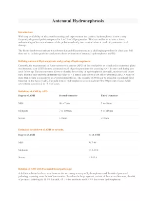
Antenatal Hydronephrosis Incidence: 1 in 1000 pregnanacy Dilatation of pelvicalyceal system ANY DILATATION EVEN IF MILD CAN NOT BE IGNORED USG is a good modality to follow & assess the severity of hydronephrosis
Natural History The outcome of ANH depends on the underlying etiology Although ANH resolves by birth or during infancy in 41-88% patients Urological abnormalities requiring intervention are identified in 4.1-15.4%
Management Monitoring of dilatation with USG Amniotic fluid should be monitored in all cases of urinary tract obstruction ???Fetal intervention Pregnancy should be carried till term unless complicated by oligohydramnios Moderate/ severe Hydronephrosis Bilateral Hydronephrosis Hydroureter Dilated Posterior urethra • • • •
Antenatal hydronephrosis Antenatal scan- 32 wks Lt hydronephrosis with dilated pelvicalyceal system; No hydroureter AP (Antero-posterior) diameter of Lt renal pelvis: 16 mm AFI: 13 How to counsel?
Counseling is Challenge….. Transient dilatation or pathological PU junction obstruction Close follow up with USG, Renal scans and MCU studies Spontaneous resolution Need of surgery and type of surgery When to intervene?
Definition of ANH by AP(Antero-Posterior) Diameter of Renal Pelvis Mild 4 to <7 mm 7 to <9 mm Moderate 7 to 10 mm 9 to 15 mm Severe >10 mm >15 mm Second trimester Third trimester
Risk Of Postnatal Pathology • Mild: 11.8% • Moderate:44.1% • Severe: 88.3%
• Isolated problem • Moderate hydronephrosis (Resolution: 40%) • CAN NOT BE IGNORED • Continue pregnancy till term • Repeat USG 48-72hrs • After delivery will require regular follow up with USG and renal scans • Need of chemoprophylaxis • Indications and type of surgery
Antenatal Hydronephrosis Rt AP of Pelvis diameter of 6 mm 20 wks scan Rt AP diameter of pelvis 8 mm 28 wks scan Rt AP diameter of pelvis 7mm 36 wks scan Before discharge Rt AP of Pelvis diameter of 7 mm Rt AP of Pelvis diameter of 5 mm USG at 1mth USG at 3mth & 1 year No dilatation
Antenatal Scan 32 wks • Bilateral hydronephrosis and hydroureter • Bilateral AP diameter 7mm • Bilateral echogenic kidneys • Bladder full; Key hole sign • AFI 8
Antenatal Scan: Hydrouretronephrosis • Vesico-ureteric reflux • Vesico-ureteric junction obstruction • Posterior Urethral Valve
Counseling • Obstruction at vesico-urethral junction • Antenatal scan at regular scans to monitor fetal growth & amniotic fluid • To continue pregnancy till term • To be investigated immediately after delivery • Need for surgery(Endoscopic Fulgaration) • Need for long term follow up & medications • Risk of ESRD in one third cases
Case… 37 wks, LSCS, 1.6 kg USG: B/L HN & HU Thinned out renal parenchyma Thickened and distended bladder Catheterised Serum Na: 132 Serum K: 5.3 S. Creatinine:1.6 VBG: Normal Urine C/S: sterile
Intestinal abnormalities HYPERECHOGENIC BOWEL • Hyperechohenic Bowel in 2nd trimester • 75% of the cases have normal outcome • Good prognosis as an isolated disorder • Needs detailed anatomical survey • Soft marker of trisomy 21 • Intrauterine CMV infection or cystic fibrosis • IUGR • Intra amniotic hemorrhage
Intestinal abnormalities Bowel Dilatation • Dilated Bowel upto 7 mm is normal • Detection rate is 40% • Echogenic bowel with dilatation • Polyhydramnios • • • Intestinal atresia Meconium ileus Meconium peritonitis
Counseling • Prognosis is good in intestinal obstruction • Need of surgery • Need of Parenteral nutrition
Tracheo-Esophageal Fistula • Difficult to diagnose • Absent stomach bubble with polyhyramnios is suggestive of pure esophageal atresia • Polyhydramnios is present in one third of patients with distal TEF • 50% of them are associated with other anomalies
Counseling Prematurity,weight and associated anomalies Surgery and complications Staged surgery Early and delayed complications Finances
Double Bubble Duodenal atresia Polyhydramnios (50%) Screening for other anomalies Cardiac (30%) Trisomy 21(30%)
Intestinal Anomalies Maturity No indication for changing the route of delivery
Cystic Lesion in abdomen Ovarian Cyst Mesenteric/ lymphatic Cyst Choledochal Cyst Prognosis good Surgery to be done after imaging
Anterior abdominal wall defects • Gastroschisis: Herniation of intestines through rt paraumbilical defect • Isolated anomaly • Elevated maternal serum alpha fetoprotein • Growth failure (30-60%), fetal demise and premature delivery • Fetal monitoring is important because of growth failure and amniotic fluid abnormalities
• Not an indication for early delivery • No indication for Cesar • Planned in set up where pediatric surgeon is readily available • Prognosis is good with survival of 90%
Omphalocele • Can be detected at 10-14 wks • 25-50% cases associated with other anomalies • Close fetal monitoring • No benefit of cesarean section • Arrangements of repair as soon as possible • May require staged repair • Survival: 70-90%
Meningomyelocele • Can be detected by 12 wks • Prognosis – Depends on level and severity/length of defect – Presence of neural tissue in sac – Severity of Presence of hydrocephalus • Closed better prognosis than open
Counseling • What Surgical treatment? • What quality of life can be given to the child? • Risk of neurological and mental deficits
• Involves enormous personal, familial and social costs • Proper counseling regarding bringing up of a handicapped child should be done • Prevention • Risk in subsequent pregnancies
Ventriculomegaly • Dilatation of lateral ventricles • Atrial diameter more than 10mm • > 15 mm is severe: Poor prognosis • Associated with neurodevelopmental problems and death
Cleft lip and Cleft Palate • 70 percent of Cleft lip/Palate & 50 percent of Cleft Palate and are non-syndromic • Other structural anomalies should be looked for especially with midline clefts • Karyotyping to be done when found to be associated with other anomalies • Diagnosed by 13-14 wks • Feeding & airway issues • Surgical timings