Download

1 / 115
1.2k likes | 1.77k Vues
Digestion &. Nutrition. 15.1 Intro to digestion. Objective Be able to describe the general functions of the digestive system. Name the major organs of the digestive system. Digestion Anatomy Gastric Secretions Accessory organs- pancreas, liver, gall bladder Small Intestine

E N D
Digestion & Nutrition
15.1 Intro to digestion Objective Be able to describe the general functions of the digestive system. Name the major organs of the digestive system.
Digestion • Anatomy • Gastric Secretions • Accessory organs- • pancreas, liver, gall bladder • Small Intestine • Large Intestine • Nutrition and Nutrients
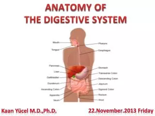
Introduction A. Digestion refers to the mechanical and chemical breakdown of foods so that nutrients can be absorbed by cells. B. The digestive system carries out the process of digestion. C. The digestive system consists of the alimentary canal, leading from mouth to anus, and several accessory organs whose secretions aid the processes of digestion.
CopyrightThe McGraw-Hill Companies, Inc. Permission required for reproduction or display.
15.2 General Characteristics of the alimentary canal Objective Describe the structure of the wall of the alimentary canal Explain how the contents of the alimentary canal are mixed and moved.
CopyrightThe McGraw-Hill Companies, Inc. Permission required for reproduction or display. General Characteristics of the Alimentary Canal A. The alimentary canal is a muscular tube that passes through the body's ventral cavity.
B. Structure of the Wall 1. The wall of the alimentary canal consists of the same four layers throughout its length, with only slight variations according to the functions of specific sections of the canal.
a. The inner layer is the mucosa, which is lined with epithelium attached to connective tissue; it protects tissues of the canal and carries on secretion and absorption.
b. The next layer is the submucosa, which is made up of loose connective tissue housing blood and lymph vessels and nerves; it nourishes the surrounding layers of the canal.
c. The muscular layer consists of inner circular fibers and outer longitudinal fibers that propel food through the canal.
d. The outer layer, or serosa, is composed of visceral peritoneum that protects underlying tissues and secretes serous fluid to keep the canal from sticking to other tissues in the abdominal cavity.
C. Movements of the Tube 1. The motor functions of the alimentary canal are of two types-- mixing movements and propelling movements. 2. Mixing movements occur when smooth muscles contract rhythmically in small sections of the tube.
3. Propelling movements include a wavelike motion called peristalsis, which is caused by contraction behind a mass of food as relaxation allows the mass to enter the next segment of the tube.
15.3 Mouth • Name the structures of the mouth and describe their functions.
D. Oral Cavity and Teeth • The mouth opens into the oral cavity • Lubricates material by mixing mucus and salivary secretions • Begins the digestion of carbohydrates and lipids Teeth 1. Two sets of teeth develop in sockets within the alveolar processes of the maxillary and mandibular bones. 2. The 20 primary teeth are shed in the order they appeared and are replaced by 32 secondary teeth. 3. Through the actions of chewing, teeth break food into smaller pieces, beginning mechanical digestion.
4. Different teeth are adapted to handle food in different ways, and include incisors, cuspids, bicuspids, and molars. 5. Each tooth consists of a crown and a root, and is made of enamel, dentin, pulp, cementum, nerves, and blood vessels. 6. A tooth is held tight in its socket by a periodontal ligament.
15.4- 15.10 Salivary Glandsand large intestines • List the enzymes the digestive organs and glands secrete, and describe the function of each enzyme. • Describe how digestive secretions are regulated. • Describe the mechanism of swallowing. • Explain how the products of digestion are absorbed.
Salivary Glands A. The salivary glands secrete saliva, which moistens and dissolves food particles, binds them together, allows tasting, helps to cleanse the mouth and teeth, and begins carbohydrate digestion.
B. Salivary Secretions 1. Salivary glands contain serous cells that produce a watery fluid with amylase, and mucous cells that produce lubricating and binding mucus. 2. Salivary glands receive parasympathetic stimulation that triggers the production of a large volume of saliva at the sight or smell of food.
C. Major Salivary Glands 1. The parotid glands, lying in front of the ear, are the largest of the major salivary glands; they secrete a clear, watery fluid rich in amylase. 2. The submandibular glands, located on the floor of the mouth, secrete a more viscous fluid. 3. The sublingual glands, inferior to the tongue, are the smallest of the major salivary glands and secrete a saliva that is thick and stringy.
15.5 Pharynx and Esophagus • Describe
Pharynx and Esophagus A. The pharynx is a cavity lying behind the mouth, and the esophagus is a muscular tube leading to the stomach.
B. Structure of the Pharynx 1. The pharynx connects the nasal and oral cavities with the larynx and esophagus and is divided into a nasopharynx (top portion), oropharynx (middle portion), and largyngopharynx (bottom portion).
C. Swallowing Mechanism 1. Swallowing reflexes can be divided into three stages. a. oral phase- Food is mixed with saliva and voluntarily forced into the pharynx with the tongue.
b. Pharyngeal phase- Sensory receptors in the pharynx sense food, which triggers swallowing reflexes. c. Esophageal phase- third stage of swallowing, peristalsis transports the food in the esophagus to the stomach.
D. Esophagus 1. The esophagus is a straight, collapsible passageway leading to the stomach. 2. Mucous glands are scattered throughout the submucosa of the esophagus and produce mucus to moisten and lubricate the inner lining of the tube. 3. The lower esophageal sphincter helps to prevent regurgitation of the stomach contents into the esophagus.
Stomach A. The stomach is a J-shaped muscular organ that receives and mixes food with digestive juices, and propels food to the small intestine.
B. Parts of the Stomach 1. The stomach is divided into cardiac, fundic, body, and pyloric regions and a pyloric canal. 2. A pyloric sphincter controls release of food from the stomach into the small intestine.
C. Gastric Secretions 1. Gastric glands within the mucosa of the stomach open as gastric pits. 2. The stomach is lined by a mucous epithelium ( an epithelium dominated by mucous cells) 3. Three types of secretory cells found in the gastric wall.
a. Mucous cells produce mucus that protects the stomach lining. b. Parietal cells secrete intrinsic factor and hydrochloric acid. Intrinsic Factor facilitates the absorption of Vitamin B12. c. Chief cells secrete pepsinogen (to digest protein) an inactive form of the enzyme pepsin. Pepsin is activated when it comes in contact with hydrochloric acid.
D. Regulation of Gastric Secretions 1. Gastric secretions are enhanced by parasympathetic impulses and the hormone gastrin, which is released from gastric glands.
2. As more food enters the small intestine, secretion of gastric juice from the stomach wall is reflexly inhibited. a. Presence of fats and proteins in the upper small intestine causes the release of cholecystokinin from the intestinal wall, which also decreases gastric mobility.
E. Gastric Absorption 1. The stomach absorbs only small quantities of water and certain salts, alcohol, and some lipid-soluble drugs.
F. Mixing and Emptying Actions 1. Following a meal, mixing actions of the stomach turn the food into chyme and pass it toward the pyloric region using peristaltic waves. 2. The rate at which the stomach empties depends on the fluidity of the chyme and the type of food.
3. As chyme fills the duodenum, stretching of its wall triggers the enterogastric reflex, which inhibits peristalysis and slows the rate at which chyme enters the small intestine.
Digestive Accessory organspancreas, liver, gall bladder Pancreas A. The pancreas has an endocrine and an exocrine function (secretion of a digestive juice called pancreatic juice).
Pancreas A. The pancreas has an exocrine function of producing pancreatic juice that aids digestion.
B. Structure of the Pancreas 1. The pancreas is closely associated with the small intestine. 2. The cells that produce pancreatic juice, called pancreatic acinar cells, make up the bulk of the pancreas.
3. Pancreatic acinar cells cluster around tiny tubes that merge to form larger ones, and then give rise to the pancreatic duct. 4. The pancreatic and bile ducts join and empty into the small intestine, which is surrounded by the hepatopancreatic sphincter.
C. Pancreatic Juice 1. Pancreatic juice contains enzymes that digest carbohydrates, fats, proteins, and nucleic acids.
2. Pancreatic enzymes include pancreatic amylase, pancreatic lipase, trypsin, chymotrypsin, carboxypeptidase, and two nucleases. 3. Protein-digesting enzymes are released in an inactive form and are activated upon reaching the small intestine.
D. Regulation of Pancreatic Secretion 1. The nervous and endocrine systems regulate release of pancreatic juice. 2. Secretin from the duodenum stimulates the release of pancreatic juice with a high bicarbonate ion concentration but few digestive enzymes.