Download
1 / 10
100 likes | 555 Vues
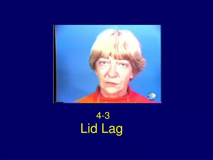
4-3. Lid Lag. Lid Lag. Von Graefe Sign Persistent elevation of the upper lid on downgaze . Classical Signs: TAO. A prominent stare. Retraction of all four eyelids Bilateral exophthalmos Hertel exophthalmometer 25 OD, 28 OS, base 108. Tight orbits/reduced orbital resilience

E N D
4-3 Lid Lag
Lid Lag Von Graefe Sign Persistent elevation of the upper lid on downgaze
Classical Signs: TAO A prominent stare. Retraction of all four eyelids Bilateral exophthalmos Hertel exophthalmometer 25 OD, 28 OS, base 108. Tight orbits/reduced orbital resilience Prominent congested scleral blood vessels A visible rim of sclera on gentle eye closure
Eye Movements Lid lag (persistent elevation of the upper eyelid in downgaze) – von Graefe sign Marked limitation of upward gaze Mild limitation of downgaze Restricted horizontal eye movements Positive forced duction test
Limited Upgaze Limitation of upgaze is due to tethering of the eyeball in the floor of the orbit by soft tissue changes. Tethering of the eyeball inferiorly can be confirmed by a forced duction test.
Limited Upgaze Duction Test • 1. Anesthetize the eye with topical anesthesia • 2. Push on the globe with a cotton tip • 3. Pull with blunt tweezers to try to move eye up. • Mechanical restriction - a positive forced duction test.
Compressive Optic Neuropathy Most serious complication Crowding of the orbital apex by enlarged ocular muscles Present in 50% severe cases TAO May require urgent orbital decompression
Figure 1 Axial CT through the orbit without contrast shows enlargement of the medial rectus muscle bilaterally. Note that the tendinous insertion is spared.
Figure 2 The coronal CT (reformatted from axial data set) without contrast shows enlargement of the medial rectus muscle, inferior rectus muscle and upper muscle complex on both sides. Courtesy of Hugh Curtin, M.D.