Download
1 / 38
380 likes | 627 Vues
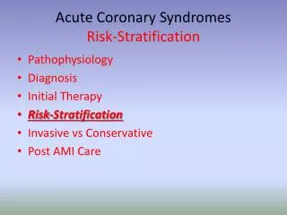
ACUTE CORONARY SYNDORME EARLY RISK STRATIFICATION. Sarah Jamison March 2003. Overview. Definition of Acute coronary syndrome (ACS) Factors used to determine risk stratification History Examination ECG changes Biochemical cardiac markers Initial management.

E N D
ACUTE CORONARY SYNDORME EARLYRISK STRATIFICATION Sarah Jamison March 2003
Overview • Definition of Acute coronary syndrome (ACS) • Factors used to determine risk stratification • History • Examination • ECG changes • Biochemical cardiac markers • Initial management
Definitions – Acute coronary syndrome • Any constellation of clinical symptoms that are compatible with acute myocardial ischemia. • It encompasses a spectrum from AMI NSTEMI UA • NSTEMI – acute process of myocardial ischemia resulting in myocardial necrosis.The initial ECG does not show ST elevation
Definitions – Acute coronary syndrome • UA – an acute process of myocardial ischemia that does not result in myocardial necrosis
Why be concerned re risk stratification……… • 1) Are the symptoms a manifestation of ACS • 2) Therapy/ site of care will vary dependent on diagnosis • 3) To determine prognosis/short term survival
History – diagnosing ACS • 5 most important factors that relate to the likelihood of ischemia due to CAD… • 1) Nature of the anginal symptoms • 2)Prior Hx of CAD • 3)Sex • 4)Age • 5)Number of traditional risk factors present • Beware – women and elderly
History – diagnosing ACS • High – Chest/L) arm pain as chief symptom,similar to previous angina Known Hx of CAD (including MI) • Intermediate – Chest/L) arm pain as chief symptom Age>70yrs/Male/Diabetes • Low– Probable ischemic symptoms in absence of any of the intermediate likelihood characteristics Recent cocaine use
History – short term risk of death or nonfatal MI in unstable angina • High – Accelerating tempo of ischemic symptoms in preceding 48hrs Pain – Prolonged ongoing (>20min) rest pain • Intermediate – Prior MI, peripheral or CVS/CABG/Aspirin use Pain – Prolonged (>20min) rest angina, now resolved, with moderate or high likelihood of CAD. Rest angina (<20min) or relieved with rest or SL NTG
History – short term risk of death or nonfatal MI in unstable angina • Low – New onset or progessive angina (Marked limitiation/or inability to carry out any physical activity) over the past 2/52. Without prolonged (>20min) rest pain but with moderate or high likelihood of CAD In patients that meet diagnostic criteria for UA/NSTEMI, the recent tempo of ischemic symptoms is the strongest predictor of risk of death
Examination - diagnosing ACS • High – Transient MR, hypotension,diaphoresis, pulmonary oedema • Intermediate – Extracardiac vascular disease • Low – Chest discomfort reproduced by palpation
Examination - short term risk of death or nonfatal MI in UA • High – Pulmonary odema, most likely secondary to ischemia New or worsening MR murmur S3 or new/worsening creps Hypotension / Bradycardia / Tachycardia Age > 75yrs • Intermediate – Age >70yrs
Examination - short term risk of death or nonfatal MI in UA Cardiogenic shock occurs in up to 5% of patients with NSTEMI and mortality rates are greater than 60%
ECG - diagnosing ACS • High – New, or presumably new, transient ST- segment deviation ( 0.05 mV) or T-wave inversion ( 0.2mV) with symptoms • Intermediate – Fixed Q waves / Abnormal ST segments or T waves not documented to be new • Low – T wave flattening or inversion in leads with dominant R waves / Normal ECG
ECG - diagnosing ACS • A completely normal ECG in a patient with chest pain DOES NOT exclude the possibility of ACS. - 1-6% of these patients it will be proven that they have had a NSTEMI - 4% will be diagnosed with unstable angina
ECG - short term risk of death or nonfatal MI in unstable angina • High – Angina at rest with transient ST-segment changes > 0.05mV Bundle – branch block, new or presumed new Sustained ventricular tachycardia • Intermediate – T wave inversion >0.2mV Pathological Q waves • Low – Normal/unchanged ECG during an episode of chest pain
ECG - short term risk of death or nonfatal MI in unstable angina • Risk factors ranked in order for risk of death in patients with ACS • 1) Confounding ECG patterns – bundle branch pattern,paced rhythm, LV hypertrophy • 2) ST segment deviation • 3) Isolated T wave inversion or normal ECG ECG pattern remains an independent predictor of death, after adjusting for clinical findings and biochemical cardiac markers
Biochemical cardiac markers • Useful in both the diagnosis of myocardial necrosis and estimation of prognosis • Prognosticaly there is a quantitative relationship between the magnitude of elevation of marker levels and the risk of an adverse event
BCM - diagnosing ACS • High – Elevated troponins or CK-MB • Intermediate – Normal • Low - Normal
A- myoglobin/CK-MB isoforms after AMI B – Cardiac Troponin after AMI C - CK-MB after AMI D – Cardiac Troponin after UA
BCM - short term risk of death or nonfatal MI in unstable angina • High– Elevated TnT > 0.1 ng/ml • Intermediate – Slightly elevated TnT (> 0.01 but <0.1 ng/ml) • Low - Normal
BCM – Creatine Kinase (CK-MB) • Advantages- Rapid, cost- efficient accurate assays. Able to detect early reinfarction • Disadvantages – Loss of specificity Low sensitivity during very early MI (6hr after sxs onset) or later after sxs onset (>36hr) and for minor myocardial damage
BCM – CK-MB isoforms • Advantages – Early detection of early MI (3-6hrs after onset of sxs) • Disadvantages – Specificity profile similar to that of CK-MB Current assays require special expertise (used predominately in research centers)
BCM - Myoglobin • Advantages – High sensitivity Useful in early detection of MI (2hrs after onset of sxs) Most useful in ruling OUT a MI • Disadvantages - Very low specificity in setting of skeletal muscle injury or disease Rapid return to normal • Should not be used in isolation
BCM – Cardiac Troponins • Advantages - Powerful tool for risk stratification Greater sensitivity and specificity than CK-MB Detection of recent onset of MI up to 2 wks after onset Useful for selection of therapy
BCM – Cardiac Troponins • Disadvantages - Low sensitivity in very early phase of MI (< 6hrs after onset of sxs) and requires repeat levels Limited ability to detect late minor reinfarction
BCM – Other markers • CRP– Patients without biochemical evidence of myocardial necrosis but who have an elevated CRP are at an increased risk of an adverse outcome • Other – Elevated levels of interleukin-6, serum amyloid A, have similar predictive value as CRP
Putting it together - management • Assign patients with chest pain to 1 of 4 groups • 1) Noncardiac • 2) Chronic stable angina • 3) Possible ACS • 4) Definite ACS
Putting it together • Most important baseline features assoc with death (Boersma et al) Age Heart rate Systolic BP ST- segment depression Signs of heart failure Elevation of cardiac markers
Putting it together • 7 point risk score (Antman et al) Age (>65yrs) More than 3 coronary risk factors Prior angiographic coronary obstruction ST – segment deviation More than 2 angina events within 24hrs Use of aspirin within 7 days Elevated cardiac markers
Summary • Risk stratification in ACS involves assessment of History Examination ECG Biochemical cardiac markers • Risk stratification is used in determining management and assessing prognosis
Summary • High risk patients – 1.7% risk of death after 30 days • Intermediate patients – 1.2% risk of death after 30 days • Low risk patients – no death after 30 days