Download
1 / 46
490 likes | 951 Vues
Neural Control and the Senses. Starr, Chapter 25. The Nervous System . The nervous system includes all the nervous tissue in the body plus the body’s sensory organs, such as the eyes and ears. The Nervous System . Nervous tissue is composed of two kinds of cells:

E N D
Neural Control and the Senses Starr, Chapter 25
The Nervous System The nervous system includes all the nervous tissue in the body plus the body’s sensory organs, such as the eyes and ears.
The Nervous System Nervous tissue is composed of two kinds of cells: • Neurons - transmit nervous system messages • Glial cells - support neurons and modify their signaling
Human Nervous System The two major divisions of the human nervous system are: • Central nervous system (CNS) • brain • spinal cord • Peripheral nervous system (PNS) • nerves that thread throughout body • plus sensory organs
Divisions of PNS • Afferent division – brings sensory info to CNS • Efferent division – carries action (motor) commands to bodies effectors – muscles and glands
Divisions of the Nervous System Central nervous system Central nervous system (CNS) information processing brain spinal cord Peripheral nervous system (PNS) sensory infor- mation travels in afferent division motor information travels in efferent division which includes... somatic nervous system autonomic nervous system sympathetic division Sensory receptors in eyes nose, etc. parasympathetic division cardiac muscle, smooth muscle glands Peripheral nervous system skeletal muscle effectors
Within PNS efferent division are two subsystems: Somatic nerves – (green) • voluntary control over skeletal muscles Autonomic nerves – (red) • involuntary regulation of smooth muscle, cardiac muscle, and glands
Autonomic system divided into: • Sympathetic division – • stimulatory effects • Respond to stress or physical activity – “fight-or-flight” response • Parasympathetic division – • relaxing effects
Opposing Systems • Most organs receive both sympathetic and parasympathetic signals • Example: Sympathetic nerves signal heart to speed up; parasympathetic stimulate it to slow down • Synaptic integration determines response
The Autonomic Nervous System Parasympathetic division (rest and digest) Sympathetic division (fight or flight) constrictspupil dilatespupil stimulatessalivation inhibitssalivation cranialnerves slowsheart acceleratesheart cervicalnerves constrictsbreathing facilitatesbreathing thoracicnerves stimulatesdigestion inhibitsdigestion stimulatesgallbladder stimulatesrelease ofglucose lumbarnerves secretesadrenaline andnoradrenaline sacralnerves contractsbladder relaxesbladder stimulatessex organs inhibits sexorgans
Sensory neurons Detect and relay info Motor neurons Transmit signals from inter-neurons to effectors Inter-neurons Receive and process info Located entirely within CNS stimulus (output) Types of Neurons receptors sensory neurons integrators interneurons of brain, spinal cord motor neurons effectors muscles, glands response (output)
Cells of the Nervous System Three types of neurons sensory neuron interneuron motor neuron afferentneuron neuron within CNS efferent neuron effector(muscle) Axon endings Anatomy of a neuron axon synapticterminals cell body dendrites Figure 27.2
Neuroglia (glial cells) A myelinated axon myelin nodes • Cells that assist, support, and protect neurons • Make up more than half the volume of the vertebrate nervous system glial cells glial cellnucleus myelincovering axon glial cellcytoplasm Anatomy of a nerve nerve bloodvessels connectivetissue axons
Nerve axon myelin sheath • A bundle of axons enclosed within a connective tissue sheath many neurons inside a connective tissue sheath
Nervous System Communication Understood easiest as a two-step process: • Signal movement down a neuron’s axon • Signal movement from this axon to second cell across structure known as synapse
interneuron motor neuron sensory neuron Information Flow • Information from sensory neurons is relayed to interneurons in spinal cord and brain • Motor neurons carry signals to body
Action Potential • How nerve cell conduct signal along axon • Inside neuron changes from negative to more positive - based on Na+ and K+ movement along membrane • Repeats from point of stimulation to move signal along membrane
Myelin Sheath • Sheath blocks ion movements • Action potential must “jump” from node to node • Greatly enhances speed of transmission
Chemical Synapse • How nerve cells send message between cells • Occurs in gap between two cells (terminal of one cell to input zone of another cell) • Neurotransmitter diffuses across synaptic cleft and binds to receptors on membrane of second cell plasma membrane of axon ending of presynaptic cell plasma membrane of postsynaptic cell synaptic vesicle synaptic cleft membrane receptor
Nervous System Communication sending cell receiving cell synapticcleft synapticterminal arrival ofnerve impulse initiation ofnew impulse mitochondrion vesicles containingneurotransmittermolecules (such asacetylcholine) neurotransmitterreceptors
The Spinal Cord Gray matter (H-shaped) • Mostly cell bodies of neurons (no myelin) White matter • Mostly axons • Sensory and motor neurons Meninges • Protective coverings
Functions of Spinal Cord • Expressway - channels sensory impulses between brain and peripheral nerves • Communication center – receives input from sensory neurons and directs motor neurons with no input from the brain • spinal impulses do not involve the brain
Reflexes • Automatic movements in response to stimuli • In simplest reflex arcs, sensory neurons synapse directly on motor neurons • Most reflexes involve an interneuron
Reflex Arc The signal from the receptor reaches asensory neuron cell body in the dorsalroot ganglion. Stimulus (tapping) arrivesand receptor is activated. afferentsignal spinalcord receptor reflexarc stimulus motorneuron efferentsignal effector The signal arrives at a sensoryneuron/motor neuron synapsein the spinal cord. Informationprocessing takes place promptinga signal to be sent through the motor neuron. The motor neuron signal stimulatesthe effector (the quadriceps muscles)to contract. Note that CNSprocessing for this reaction washandled entirely in the spinal cord;the brain was not involved. response
The Human Brain • There are six major regions in the adult brain: • Cerebrum • Thalamus and hypothalamus • Midbrain • Pons • Cerebellum • Medulla oblongata
The Vertebrate Brain* corpus callosum hypothalamus thalamus pineal gland location part of optic nerve midbrain cerebellum pons medulla oblongata Fig. 25-15, p.434
Cerebrospinal Fluid • Surrounds the spinal cord • Fills ventricles within the brain • Blood-brain barrier controls which solutes enter the cerebrospinal fluid
Anatomy of the Cerebrum • Largest and most complex part of human brain • Divided into right and left cerebral hemispheres • Thin outer layer (cerebral cortex) is site of our highest thinking
Lobes of the Cerebrum primary somatosensory cortex primary motor cortex parietal frontal occipital temporal
The Human Brain • The brainstem is a collective term for three brain areas—the midbrain, pons, and medulla oblongata • These brainstem structures are active in: • Controlling involuntary bodily activities (such as breathing and digesting). • Relaying information. • Processing sensory information.
The Brain Stem cerebralcortex cerebrum cerebellum thalamus hypothalamus pituitarygland midbrain pons brainstem medullaoblongata Figure 27.9
The Human Brain • Most of the body’s sensory perceptions are channeled through the thalamus before going to the cerebral cortex. • The hypothalamus is important in sensing internal conditions and in maintaining stability or homeostasis in the body, largely through its control of many of the body’s hormones.
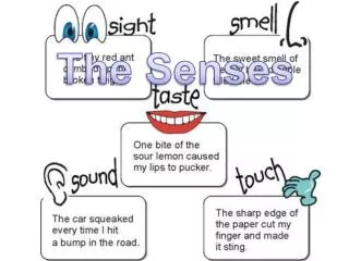
Our Senses Each sense employs cells called sensory receptors that do two things: • Respond to stimuli • Transform these responses into the language of the nervous system – electrical signals that travel through action potentials
Smell • A special sense • Olfactory receptors • Receptor axons lead to olfactory lobe olfactory bulb receptor cell
Taste • A special sense • Chemoreceptors • Five primary sensations: • sweet, sour, salty, bitter, and umami
Our Sense of Vision • Perceives visual field • Lens collects light • Image formed on retina
Human Eye sclera retina choroid iris fovea optic disk lens pupil cornea part of optic nerve aqueous humor ciliary muscle vitreous body
Pattern of Stimulation • Image on retina is upside down and reversed right to left compared with the stimulus • Brain corrects during processing
Retinal Stimulation Patterns a Light rays from an object converge on the retina, form an inverted, reversed image. muscle contracted b When a ciliary muscle contracts, the lens bulges, bending the light rays from a close object so that they become focused on the retina. close object slack fibers c When the muscle relaxes, the lens flattens, focusing light rays from adistant object on the retina. muscle relaxed distant object taut fibers Fig. 25-26, p.442
Organization of Retina • Photoreceptors at back of retina, in front of pigmented epithelium • For light to reach photoreceptors, it must pass layers of neurons involved in visual processing
Organization of Retina • Signals from photoreceptors are passed to bipolar sensory neurons, then to ganglion cells • Axons of ganglion cells form the two optic nerves Cone Rod Bipolar sensory neuron Ganglion cell
The Photoreceptors • Rods • Contain the pigment rhodopsin • Detect very dim light, changes in light intensity • Cones • Three kinds; detect red, blue, or green • Provide color sense and daytime vision
Hearing • Outer ear • Middle ear • Inner ear