Download
1 / 40
450 likes | 1.31k Vues
Histiocytic Syndromes. Dendritic cell-related disorders Langerhans cells histiocytosis juvenile xanthogranuloma Macrophage-related disorders primary hemophagocytic syndromes (familial, sporadic) secondary hemophagocytic syndromes (viral, other) Malignant disorders monocytic leukemias

E N D
Histiocytic Syndromes • Dendritic cell-related disorders • Langerhans cells histiocytosis • juvenile xanthogranuloma • Macrophage-related disorders • primary hemophagocytic syndromes (familial, sporadic) • secondary hemophagocytic syndromes (viral, other) • Malignant disorders • monocytic leukemias • malignant histiocytosis
Langerhans Cell Histiocytosis Epidemiology • Incidence: • 5 cases/million population/yr (childhood LCH) • incidence of adult LCH is unknown • more common in males than females (1.3 : 1) • average age at onset: 1.8 years
Langerhans Cell Histiocytosis: Historical Syndromes • eosinophilic granuloma: isolated lytic lesion of bone or soft tissue mass • Hand-Schuller-Christian disease: skull lesions, exophthalmos, and DI (1893) • Letterer-Siwe disease: seborrheic rash, hepatosplenomegaly, lung involvement in infants (1924) • the spectrum of “Histiocytosis X” (1953)
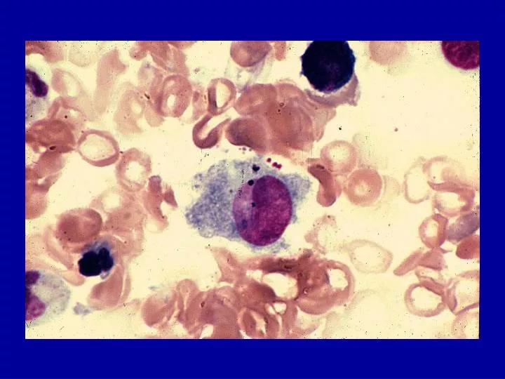
Langerhans cells • described by Paul Langerhans (medical student) in 1868 • Langerhans cells (LC) reside in the epidermis (1-2% epidermal cells) & lung • LCs process antigens for presentation to T cells after migrating to lymph nodes • LCs contain Birbeck granules, and express CD1a and S-100 • LCs associated with “Histiocytosis X” in 1973 (Nezelof et al)
Generation of Langerhans cells • LCs can be generated from stem cells: • obtain CD34+ stem cells • incubate w/ GM-CSF and TNF-alpha for 3 weeks • resulting cells have Birbeck granules and express CD1a • have functional characteristics of LC’s
Langerhans Cell Histiocytosis: Pathology • a clonal proliferation of Langerhans cells • LCs do not show dysplasia or atypia (features of malignant cells) • LCH lesions contain LCs, macrophages, T cells, eosinophils, and granulocytes • LCs associated with “Histiocytosis X” in 1973 (Nezelof et al)
Langerhans Cell Histiocytosis: Clinical manifestations I • painful swelling of bones • unifocal bony lesion (31% at presentation) • isolated multifocal bone involvement (19%) • persistent otitis / mastoiditis • mandible involvement (“floating teeth”) • papular rash (37% at presentation) • hepatosplenomegaly • lymphadenopathy
Langerhans Cell Histiocytosis: Clinical manifestations II • pulmonary involvement (interstitial pattern -> “honeycombing”) • marrow involvement (cytopenias) • GI involvement (diarrhea, malabsorption) • endocrine involvement: • diabetes insipidus • growth failure • hypothyroidism
Langerhans Cell Histiocytosis: Extent of disease at diagnosis • single system / single site 33% • single system / multiple sites 15% • multisystem involvement 52%
Langerhans Cell Histiocytosis: Diagnostic criteria (Histiocyte Society, 1987) • Presumptive diagnosis • light morphology • Designated diagnosis • light morphology, plus • two or more positive stains for ATPase, S-100, a-D-mannosidase, peanut lectin • Definitive diagnosis • light morphology, plus • Birbeck granules and/or CD1a staining
Langerhans Cell Histiocytosis: Natural history • isolated skin involvement (“Hashimoto-Pritzker disease”): spontaneous resolution • eosinophilic granuloma: may resolve or progress; responds to biopsy, curettage • Hand-Schuller-Christian disease: usually fatal if untreated due to DI • Letterer-Siwe disease: usually fatal if untreated
Langerhans Cell Histiocytosis: Therapeutic modalities • biopsy or curettage • radiation therapy (low dose) • topical steroids • intralesional steroid injections • oral or intravenous steroids • oral or intravenous chemotherapy • single agents (vinblastine, etoposide) • combination chemotherapy
LCH-I: Design • first international clinical trial for LCH • compared vinblastine vs etoposide when given with steroids • enrolled 447 pts from 1991-1995 • 143 randomized pts with multisystem disease
DAL HX-83 and HX-90 studies: Design • two multi-center, non-randomized trials in Austria, Germany, Netherlands and Switzerland • risk-adapted assignment to treatment • intensive induction and continuation therapy (much like leukemia therapy) • total duration of therapy was 12 months
LCH-II • compared vinblastine/prednisone +/- etoposide as induction therapy • continuation therapy: 6-MP, with pulses of induction therapy agents • total duration of therapy was 24 weeks • enrolled 697 pts from 1996-2000 • stratified patients on basis of risk
LCH-II: Risk stratification • “Risk” patients: involvement of liver, spleen, lungs, bone marrow; age < 2 yrs • “Low-risk” patients: none of the above
DAL HX, LCH-I and LCH-II: Conclusions • overall survival of multi-system patients was about 80% on all studies • patients with lack of response at week 12 have a high risk of poor outcome • 20% of patients do not respond to current therapy -> new treatments needed • prolonged therapy has potential benefit
LCH-III: Overall Goals • to deliver risk-adapted therapy • to evaluate response in various risk groups • to assess morbidity in various risk groups
LCH-III: Design • adds methotrexate for “risk” patients • adds stratifications for multifocal bone only patients and CNS patients • patients with involvement of facial bones or middle cranial fossa have 3-fold risk for DI
LCH in Adults • most adults with LCH have pulmonary LCH • most are smokers • symptoms: cough, shortness of breath, chest pain, sputum production, pneumothoraces • CXR: diffuse bilateral infiltrates -> progress to cyst formation and “honeycombing” • Treatment: reports that 2-CdA is effective
Adults Some lesions are not clonal LC cells more mature: CD86+ No IL-10 in macrophages Children All lesions are clonal LC cells less mature: CD86- IL-10 expressed in macrophages LCHChildren vs Adults
Treatment Options for Recurrent/Refractory LCH • Other chemo (Ara-C, methotrexate, cytoxan) • cyclosporine • interferon • retinoic acid (France) • thalidomide (Texas Children’s Cancer Ctr) • allogeneic bone marrow transplantation • 2-chlorodeoxyadenosine (2-CdA) +/- Ara-C
2-CdA for refractory LCH • Review: 27 pts with refractory LCH were treated with 2CdA (23) or 2-DCF (4) • Doses: 0.1 mg/kg/d - 13 mg/m2/day x 5-7 days for 1-6 courses • Results: 15 CR, 5 PR, 5 NR; no toxic deaths • Toxicities: myelosuppression, prolonged thrombocytopenia, peripheral neuropathies
LCH-S-98: Salvage trial • for pts with relapsed or refractory LCH • must have failed multi-agent therapy and have high-risk disease • 2-CdA 5 mg/m2/day x 5 days q 3 wks x 6 courses • next salvage trial will add low-dose Ara-C to this dose of 2-CdA
Langerhans Cell Histiocytosis: Why does it happen? • Epidemiologic study of possible risk factors published in 1997 • conducted in conjunction with HAA • 22-page self-administered questionnaire • parents of 900 LCH patients in HAA • 63% response rate • 459 patients met all eligibility criteria
Langerhans Cell Histiocytosis: Study of risk factors • Possible associations: • neonatal infections (cause or effect?) • exposure to solvents (acetone) • thyroid disease in family members • No association: • in utero exposure to cigarette smoke • maternal infections or medications
Langerhans Cell Histiocytosis: Challenges for the Future • Better understanding of histiocyte biology • differences between normal LC’s, LCH • differences between localized, extensive LCH • differences between childhood, adult LCH • Better understanding of LCH epidemiology • genetic and environmental factors
Langerhans Cell Histiocytosis: Challenges for the Future • New treatments for both newly diagnosed and relapsed patients • more effective • fewer side effects • “targeted” therapy (CD1a-linked radioisotope)