Download
1 / 81
980 likes | 2.48k Vues
GYNECOLOGICAL EXAM. SFC WARD. Do a Complete Physical Assessment. HEENT CV Lungs Breasts Abdomen Pelvic/rectal Neuro Musculoskeletal. Essentials for an Adequate Examination--Relaxation.

E N D
GYNECOLOGICAL EXAM SFC WARD
Do a Complete Physical Assessment • HEENT • CV • Lungs • Breasts • Abdomen • Pelvic/rectal • Neuro • Musculoskeletal
Essentials for an Adequate Examination--Relaxation • Patient should be given an opportunity to empty her bladder prior to the exam-- Routine UA specimen may be obtained at this time • Explain what is to take place during the exam • Drape her appropriately, cover extending at least over her knees • Arms should be at her side or folded across her chest.
Essentials for an Adequate Examination • Examiner's hands should be warmed, also warm the speculum before the exam • Have eye to eye contact with the patient during the exam • Explain in advance each step in the examination, avoiding any sudden or unexpected movements
Correct Examining Position of the Patient • The Lithotomy Position/or Semi-Sitting Lithotomy Position • Lying in supine position • Thighs flexed and abducted • Feet resting in stirrups • Buttocks extended slightly beyond edge of exam table • Head supported with a pillow
Male examiners should always be attended by female assistants • Hx should be taken prior to patient disrobing. • Do not enter the room with an unclothed patient unless you have a female chaperone.
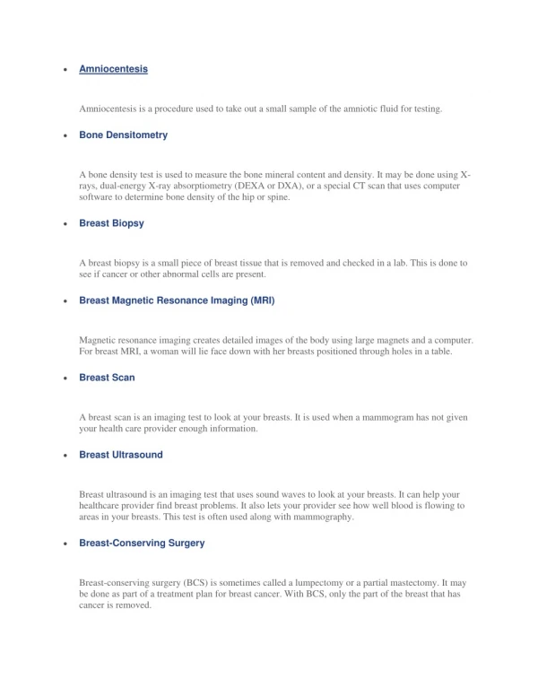
Breast Examination (note the following): • Breast development • Size, symmetry, contour and appearance of the skin (Variation in breast contour may include the presence of masses, dimpling, or flattening.)
Breast Examination (note the following): • Nipples • Direction of the nipples may provide a clue to masses when there is asymmetry • Discharge may indicate disease or may merely occur with the hormonal fluctuation of the menstrual cycle • Ulcerated areas and other nipple lesions require further exploration • Lymph node, have patient press hands against hip contracting pectoral muscles, palpate lateral group of axillary lymph nodes
Nipple Laceration Paget’s Disease Inverted Nipple
Sequence of Breast Exam • Patient sitting or standing--press hands on hips to contract pectoral muscles (This maneuver accentuates any existing tissue retraction.) • Observe size and contour and appearance of the skin • Observe direction of nipples
Sequence of Breast Exam • Palpate axillary region
Sequence of Breast Exam • Have patient lying flat with arm abducted and hand under head (This helps flatten breast tissue evenly over the chest wall.) • Palpate entire breast and lymph nodes, (axillary and infraclavicular) • Palpation is performed in a rotary motion using an organized approach
Sequence of Breast Exam • Some examiners start in the upper outer quadrant where tumors develop most frequently • Breasts of young clients are firm and elastic • Older clients, the tissue may feel stringy and nodular.
Sequence of Breast Exam • Palpating large pendulous breasts, use a bimanual technique. The inferior portion of the breast is supported in one hand while the other hand palpates breast tissue against the supporting hand • Special attention is given to palpate the nipples, and areola • Entire surface is gently palpated • With thumb and index finger compress the nipple; note any discharge.
NOTE: If client complains of a mass or tenderness of one breast, examine the opposite breast first to ensure an objective comparison of normal and abnormal tissue.
Sequence of a Pelvic Examination • Inspect the client's external genitalia • Perineal area must be well illuminated • Both hands are gloved to prevent the spread of infection • Perineum is sensitive and tender, warn the client by touching the neighboring thigh first before advancing to the perineum.
NOTE: A client suffering pain or deformity of the joints may be unable to assume a Lithotomy position. It may be necessary to have the client abduct only one leg or have another person assist in separating the client's thighs.
Sequence of a Pelvic Examination • Mons pubis--note quantity and distribution of hair growth • Labia--usually plump and well-formed in adult female • Perineum--slightly darker than the skin of the rest of the body. Mucous membranes appear dark pink and moist
Sequence of a Pelvic Examination • Separate the labia and inspect the labia minora: • Labia minora • Clitoris • Urethral orifice • Hymen • Vaginal orifice
Sequence of a Pelvic Examination • Note the following: • Discharge • Inflammation • Edema • Ulceration • Lesions
Sequence of a Pelvic Examination • Note abnormalities such as: • Bulges and swelling of vulva and vagina • Enlarged clitoris • Syphilitic chancres • Sebaceous cyst Primary Syphilis
Sequence of a Pelvic Examination • Skene's glands • Near the urethra • Suspect inflammation; check for urethral discharge (Dc = Infxn Most likely GC) • Insert index finger with palm facing you into the vagina up to the 2d joint. Apply pressure upwards and milk the Skene's gland by moving your fingers outward • Do this on both sides and note COCA on any discharge. Obtain specimen for culture. • Change glove if discharge is found.
Sequence of a Pelvic Examination • If there is history or appearance of labial swelling check Bartholin's glands • Insert index finger up to first knuckle • With your index finger and thumb, palpate the posterolateral area of the labia majora noting any: • Swelling • Tenderness • Masses • Heat or discharge
Sequence of a Pelvic Examination • Bartholin's glands (CONT) • A painful abscess is pus filled and usually staphylococcal or gonococcal in origin and should be incised and drained to perform C+S.
Sequence of a Pelvic Examination • Assess the support of the vaginal outlet: • With the labia separated by middle and index finger • Ask patient to strain down • Note any bulging of the vaginal walls (cystocele and rectocele).
Sequence of a Pelvic Examination • Inspect the anus at this time, note presence of lesions and hemorrhoids
Speculum Examination of Internal Genitalia • Select a speculum of appropriate size, lubricate and warm with warm water (Commercially prepared lubricants interfere with pap smear studies) • Small--not sexually active female • Medium--sexually active • Large--women who have had children • Medium to large speculum may be used if female has had children.
Speculum Examination of Internal Genitalia • Hold speculum in right hand • Place two fingers just inside or at the introitus and gently press down, this will help guide the speculum into the vagina opening • The speculum has to be closed • Insert closed speculum obliquely into vagina at a 45 degree angle rotating 50 degrees counterclockwise
Speculum Examination of Internal Genitalia • Avoid trauma to the urethra • Care is taken to avoid pulling pubic hair or pinching the labia • Maintaining downward pressure, open blades slowly after full insertion and position the speculum so that the cervix can be visualized • When the cervix is in full view, the blades are locked in the open position
Examination/Collection Specimen of the Cervix • Inspect the cervix • Color should be uniformly pink • Erythema around os: • Ectropion--expressed columnar epithelium • Erosion--term has been used to describe both the exposed columnar epithelium and the erythema seen with cervicitis • Pale--anemia • Bluish--Chadwick's sign, presumptive sign of pregnancy.
Examination/Collection Specimen of the Cervix • Inspect the cervix • Lesions/cysts: • Nabothian cyst--endocervical retention cysts usually secondary to cervical infection/inflammation • Friable, granular, red or white patchy areas--be suspicious of dysplasia, needs to be evaluated with colposcopy • Ulcerative lesions--may be herpetic; do viral culture of lesions and refer for colposcopy • Polyps--soft, friable mass protruding through os; may bleed if traumatized; refer for eval/removal
Examination/Collection Specimen of the Cervix • Inspect the cervix • Discharge: • Endocervical vs. from vaginal vault • Physiological discharge--odorless, colorless • Culture any discharge.
Examination/Collection Specimen of the Cervix • Inspect the cervix • Os: • Nulliparous--small, round, oval • Parous/multiparous--linear, irregular, stellate
Examination/Collection Specimen of the Cervix • Obtain specimens • Chlamydia culture--most prevalent STD • GC culture--gram stain not reliable, done for screening, must do Thayer-Martin for confirmation • PAP smear for cytology--sites of collection: • Endocervical brush--all patients • Endocervical scrape with spatula--all patients • Posterior fornix--all • Vaginal cuff and area of former posterior fornix for post-hysterectomy patient.
Examination/Collection Specimen of the Cervix • Obtain specimens • Wet mount of normal saline: • WBCs--evidence of infection/inflammatory process • Flagellated trichomonads--trichomonas • Granulated epithelial cells,"clue cells"--Gardnerella
Examination/Collection Specimen of the Cervix • Obtain specimens • KOH prep--budding yeast--candidiasis + "whiff" (fishy odor)--Gardnerella • Viral cultures of suspected lesions • Others: • STS (RPR/VDRL)--if suspected STDs • Beta HCG--if pregnancy suspected.
Examination/Collection Specimen of the Cervix • Obtain specimens • Collect during routine PAP smear/pelvic exam: • Wet mount if suspicious discharge • KOH prep if suspicious discharge • Thayer-Martin of Transgrow cultures • Chlamydia cultures
Inspection of the Vagina • Withdraw the speculum slowly while observing the vaginal wall • Close blades as the speculum emerges from the introitus • Inspect vaginal mucosa as the speculum is withdrawn