Download
1 / 19
190 likes | 506 Vues
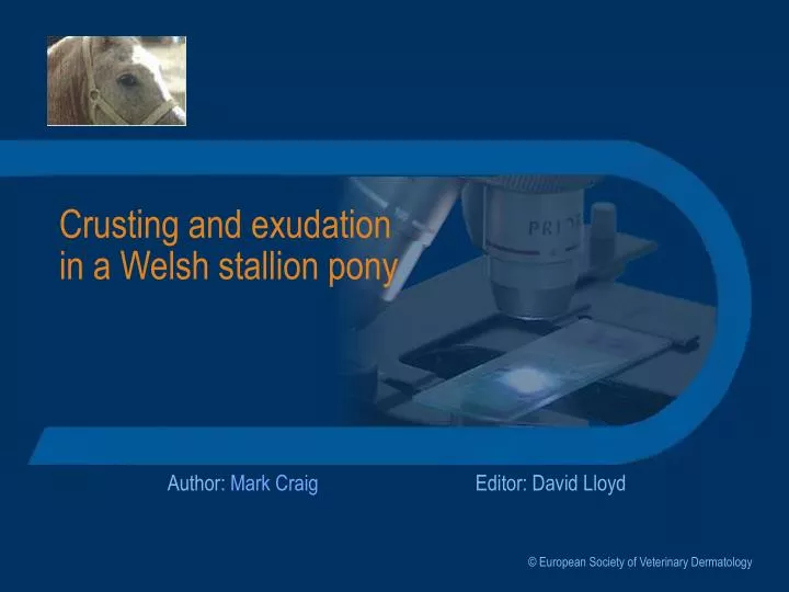
Crusting and exudation in a Welsh stallion pony. Author: Mark Craig. Editor: David Lloyd. © European Society of Veterinary Dermatology. History -1. 10-year-old Welsh pony stallion Weight 300 kg . Click to reveal the text on this screen Click the forward arrow to jump to the next screen.

E N D
Crusting and exudation in a Welsh stallion pony Author: Mark Craig Editor: David Lloyd © European Society of Veterinary Dermatology
History -1 • 10-year-old Welsh pony stallion • Weight 300 kg Click to reveal the text on this screen Click the forward arrow to jump to the next screen History
History -2 • First signs developing over a 4-week period • Papules on flank and neck, pruritus • Generalised crusting and exudation • Swelling of all four legs accompanied by stiffness and lameness • Weight loss and lethargy • No treatment by the referring vet History
Clinical signs - 1 The horse was thin and there was generalised crusting with diffuse alopecia No peripheral lymphadenopathy was detected Signs
Clinical signs - 2 Close-up views of alopecic andcrusted areas Clipped area on the withers Signs
How would youapproach this case? • What are the next steps you would take? • Make a list of your principle differential diagnoses • List any samples you would collect • List any tests you would perform to assist in making a definitive diagnosis Signs
Test - 1 • Principle differential diagnoses • Bacterial folliculitis, dermatophilosis, dermatophytosis • Ectoparasitic infestation • Pemphigus complex, SLE, drug eruption • Allergy Differentials
Tests - 2 • Tests • Blood tests: routine haematology and biochemical screens; ANA test • Multiple punch and excision biopsy samples from crusted and alopecic areas • Skin scrapings, crusts and hair pluckings for microscopic examination, smears, bacterial and fungal culture Tests
Results • Scrapings, crusts and hairs did not reveal ectoparasites or fungal structures; Dermatophilus was not demonstrated on microscopic examination • Haematological and biochemical profiles were within accepted limits. The ANA test was negative • Histopathology revealed a superficial pustular dermatitis. No micro-organisms were found in the lesions Tests
What now? • What treatment, if any, should you now institute whilst waiting for the fungal cultures? • What are now your principle differential diagnoses? • Are there any other samples you would collect? Tests
Initial therapy • Ectoparasitism and neoplasia were deemed unlikely • Autoimmune or immune-mediated disease seemed likely and the horse was deteriorating hence therapy was initiated • Prednisolone, 0.5 mg/kg daily; trimethoprim and sulphadiazine (Uniprim), 0.5 sachet daily Therapy
What is yourdiagnosis? • What is your principle diagnosis? • Do the investigations permit a definitive diagnosis? • Are there any additional investigations which you think may need to be done? Therapy
Histopathology Further tests • A superficial pustular dermatitis was present • The pustules contained many neutrophils & acanthocytes but no micro-organisms Therapy
Diagnosis • Pemphigus foliaceus • Lesion type and histopathology are consistent • No history of previous drug therapy • Fungal culture was negative Therapy
How would you deal with this case? • What is your prognosis? • How will you advise the owner? • What treatment would you consider? Therapy
Response to therapy - 1 • After 5 days the lesions were unchanged • The prednisolone dose was increased to 300 mg per day and there was a moderate improvement over a period of 10 days - decreased scaling and some hair regrowth • Steroid therapy lapsed (owner’s choice) and the horse’s condition remained stable over a two-month period but then again worsened Notes
Response to therapy - 2 • Prednisolone therapy at 300 mg twice daily (1 mg/kg b.i.d.) was resumed. Within 2 weeks all lesions had disappeared and after 1 month the horse was reported to be in good condition • Attempts will be made to lower the dose over the coming months Notes
Prognosis • Prognosis is guarded • The disease may require potent immunosuppressive therapy with significant side effects • It may not respond adequately Notes
Review If you would like to review this case, please use the navigation buttons below Notes