Download
1 / 27
270 likes | 621 Vues
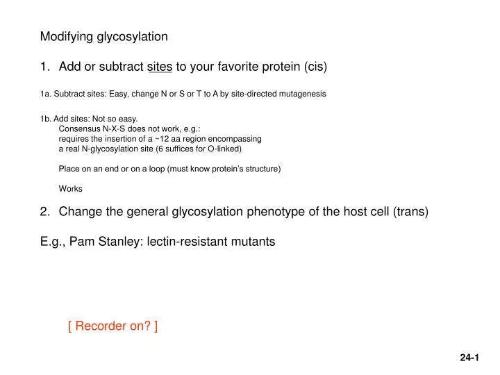
Modifying glycosylation Add or subtract sites to your favorite protein (cis) 1a. Subtract sites: Easy, change N or S or T to A by site-directed mutagenesis 1b. Add sites: Not so easy. Consensus N-X-S does not work, e.g.: requires the insertion of a ~12 aa region encompassing

E N D
Modifying glycosylation • Add or subtract sites to your favorite protein (cis) • 1a. Subtract sites: Easy, change N or S or T to A by site-directed mutagenesis • 1b. Add sites: Not so easy. • Consensus N-X-S does not work, e.g.: • requires the insertion of a ~12 aa region encompassing • a real N-glycosylation site (6 suffices for O-linked) • Place on an end or on a loop (must know protein’s structure) • Works • Change the general glycosylation phenotype of the host cell (trans) • E.g., Pam Stanley: lectin-resistant mutants [ Recorder on? ]
Modifying glycosylation • Add or subtract sites to your favorite protein (cis) • Change the general glycosylation phenotype of the host cell (trans) Clone enzyme genes:Glycosyl transferases, mostlyAlso some synthetases (e.g., NAcNeu, i.e., biosynthetic path to sialic acid) Can be complex:e.g., 7 different fucosyl transferases (FTs),with different (overlapping) substrate specificities Simpler example: Hamster cells do only 2,3 sialylation. Humans do 2,6 as well, via a 2,6 sialyl transferase (ST) Experiment:Over-express cloned human 2,6 ST, along with a substrate protein.producing permanent transfectants of CHO cells Works: Get both types of structures now, substantially (although not exactly the same ratio as in human cells). Zhang, X., Lok, S.H., and Kon, O.L. 1998. Stable expression of human alpha-2,6-sialyltransferase in Chinese hamster ovary cells: functional consequences for human erythropoietin expression and bioactivity. Biochim Biophys Acta1425: 441-452.
Isolate mutant mammalian cell lines deficient in specific glycosylation enzymes Stanley: Isolation of multiply mutated glycosylation mutants by selecting for lectin resistance Lectins = carbohydrate-binding proteins Plant lectins used mostly here (but occur widely in animals as well) Sequential selections, push - pull on resistance, sensitivity Resistance: enzyme deficiency failure to add the sugar need for lectin binding Increased sensitivity: failure to add a sugar produces greater exposure of underlying sugars in a transferase-negative mutant better binding to the exposed sugar Showed power of selection Showed usefulness of complementation analysis via cell hybridization Hybrid selection: All lec-R mutants were: WGA (wheat germ agglutinin) resistant (various degrees) & pro- Tester parent was single lec-R + GAT- (req. glycine, adenine and thymidine) Select in medium lacking pro, GAT, and with +/- WGA Complementing hybrids will have regained sensitivity to WGA Mutants in the same gene will remain WGA resistant (non-complementation) Could now be used as a tabla rasa (blank slate) for the introduction of a series of enzymes to build custom-tailored glycoconjugates. Complicated though (order of addition, location in the Golgi, etc. ) Potential: targeting to carbohydrate-sensitive receptors (e.g., liver asialoglycoprotein receptor); clearance rate Pam Stanley Review: Nature Biotechnology 19, 913 - 917 (2001) , The bittersweet promise of glycobiology. Alan Dove
Umana, P., Jean-Mairet, J., Moudry, R., Amstutz, H., and Bailey, J.E. 1999. Engineered glycoforms of an antineuroblastoma IgG1 with optimized antibody-dependent cellular cytotoxic activity. Nat Biotechnol17: 176-180. Target here (bisecting NAcGlc) (NAcG = N-acetyl-glucosamine here) Presence of the bisecting NAcGlc enhances binding of T-cell receptor to the Fc region of antibodies. Binding is needed for ADCC. Mouse and hamster cell lines used for commercial production lack the glycosyltransferase needed for bisecting NAcG addition A rat myeloma cell line does produce MAb with the bisecting NAcG. Hypothesis: Expression of the rat enzyme in a CHO cell line will add a bisecting NacG to the anti-neuroblastoma MAb produced by these cells. The modified MAb will be a better mediator of ADCC. Experiment: Clone the cDNA for this enzyme from the rat line and transfer it to CHO cells, driven by an inducible tet promoter. Check sugar structure of MAb and ADCC efficiency of the MAb. Both improved.
TARGET CELL (Killer T-cell) Genentech Commercial MAb injected as a therapeutic T-cell surface receptor binds Fc region of antibody molecule(Fc gammaR)
Protein Glycosylation Knock-out Assigned: Naoko Yamane-Ohnuki, et al.. Establishment of FUT8 knockout Chinese hamster ovary cells: an ideal host cell line for producing completely defucosylated antibodies with enhanced antibody-dependent cellular cytotoxicity. Biotechnol Bioeng. 2004 Sep 5;87(5):614-22 Optional Update: Kanda Y, Yamane-Ohnuki N, Sakai N, Yamano K, Nakano R, Inoue M, Misaka H, Iida S, Wakitani M, Konno Y, Yano K, Shitara K, Hosoi S, Satoh M. Comparison of cell lines for stable production of fucose-negative antibodies with enhanced ADCC. Biotechnol Bioeng. 2006 Jul 5;94(4):680-8. Other refs. Review: Grabenhorst, E., Schlenke, P., Pohl,., Nimtz, M., and Conradt, H.S. 1999. Genetic engineering of recombinant glycoproteins and the glycosylation pathway in mammalian host cells. Glycoconj J16: 81-97. Background: Stanley, P. 1989. Chinese hamster ovary cell mutants with multiple glycosylation defects for production of glycoproteins with minimal carbohydrate heterogeneity. Mol Cell Biol9: 377-383.
Biotechnol Bioeng. 2004 Sep 5;87(5):614-22 Hypothesis: Fucose interferes with binding of the T-cell Fc-gamma-3 receptor to the Fc region of an antibody molecule. Elimination of fucose from produced MAbs will increase ADCC Create a mutant CHO cells (starting with amplifiable dhfr- cells) in which the fucose trasnferase genes have been knocked out. All MAbs produced in these mutant cells will be better at promoting ADCC
Double knock-out strategy for FUT8 an alpha-1,6,fucosyl transferase Little sequence data available for Chinese hamster Isolate CHO cDNA using mouse sequence data for primers Use CHO cDNA to isolate CHO genomic fragments from a commercial lambda library K.O. exon 1 translation start region Homology regions DT= diphtheria toxin gene, Kills if integrated via non-homologous recombination For hemizygote: Select for G418 resistance, Screen by PCR for homologous recomb. 108 cells 45000 colonies 40 false recombinants (extension-duplications) + 1 true recombinant Step 2 for homozygote, select for Pur-resistance 1.6X10870,000 screened 10 double KO homozygotes. Lox sites for later removal of drug resistance marker Remove drug resis. genes by transient transfection with Cre Recombinase. Exon 1 has undergone a 200 nt deletion. Note: 10’s of thousands of PCRs performed to screen for homologous recomb., using 96-well plates
Double knockout evidence After Cre treatment Original KO’d genes have a 1.5 kb insertion (Southern blot) Final mRNA has 200 nt deletion (RT-PCR
Use of a fluoresceinated lentil lectin (LCA) that binds fucose oligosaccharides to demonstrate lack of fucosylation in glycosylated proteins in the FUT8 -/- cells Control background fluorescence(FL-anti avidin) FUT8 +/+ FUT8 +/- Surprising that fucosylation levelis down by one-half; that CHO cells do not have excess fucosylation capacity . . . FUT8 -/-
Rituxan (anti-CD20) produced in FUT -/- cells does not contain fucose (CD20 is expressed at a high level on the surface of many lymphoma cells.) HPLC analysis Digestion all the way to monosaccharides Missing d - g
Binding to CD20 membranes is the same FUT8-/- anti CD20 = Ritxuan In ADCC, FUT8-/- anti-CD20 >> Rituxan Anti-CD20 from a partially FUT-deficient rat cell line Characteristics of“FUT8-/- anti-CD20” Fc-Receptor protein binding assay Rat line FUT-/-’s Complement-mediated cell toxicity is the same for FUT8-/- and Rituxan(not T-cell mediated ADCC) Rituxan = commercial product, 98% fucosylated
Very laborious, but apparently a big payoff. Better selection?: Why not use the fluorescent LCA to select for the FUT8 KO’s along with G418 resistance (double, sequential selection) ? Avoid 100,000 PCRs. . .
Second generation protein therapeutics: improvements over nature. Or: Isolation of recombinant protein mutants with altered binding properties. Here: Tissue plasminogen activator (TPA, clot-buster) Why are they doing this? Problem: Binding of TPA to liver cells leads to clearance from the bloodstream Want to avoid clearance in TPA therapy (anti-thrombolytic, clot buster) Know: MAb387 blocks binding to cultured hepatoma cells (liver-like) Know: MAb387 decreases clearance rate. Goal: Mutate the cloned TPA gene. Mutate it in the MAb387-binding region mutant TPA that: 1) is hepatocyte binding-negative (select) 2) is still protease+ (remains catalytically active) (screen)
How could one do this? Select? Need to characterize many mutant proteins, and find the protein with the desired characteristic, and then rescue the gene for that protein. Mammalian cell transfectants: But TPA is secreted, so protein becomes divorced from the DNA that coded it. My editorializing: But Coffino and Scharff had a technique for looking at secretory variants (of myeloma cells): Immunoprecipitate secreted proteins around colonies grown in agar (Ig secretion, precipitation by goat anti-mouse-Ig antibody):
All in soft agar Medium in agar Imagine: Antibody in top layer = MAb387 Colonies = CHO cell permanent co-transfectants of mutant library TPA Longer. Colonies may not make enough. But you don’t need a FACS ($$) Precipitate - Precipitate + Coffino and Scharff (Proc Natl Acad Sci U S A. 1971 Jan;68(1):219-23.)
Consider an alternative: Phage display: a way to link the variant protein to its coding DNA Mutagenize the gene as a fusion protein to a phage coat protein and make a library in bacteriophage. The mutants will be displayed on the surface of the phage and can be “panned” for (or against). DNA DNA DNA Here, one would collect the members of the phage library of mutant TPAsthat don’t stick to hepatoma cells, or to immobilized MAb387. But lots of noise in a negative selection = non-stickers could be for many uninteresting reasons (denatured, statistical, etc. )
Authors here use a mammalian cells as the carrier of the DNA and the cell surface as a display site. Via making a fusion protein to a membrane anchor protein, DAF (peptide, really). What did they do? Mutagenesis: What region? 333 bp K1 (kringle-1), known to bind the MAb387, which competes for hepatocyte binding (so assuming it is the same target epitope). How did they get it mutated? Error-prone PCR How did they isolate just the kringle 1 region? PCR fragment. How did they get the mutagenized fragment back in? Introduced restriction sites at the ends, w/o affecting the coding.
What did they put the mutagenized fragment into? DAF – TPA fusion protein geneHow did they get it into into cells? Electroporation How many copies per cell. And why is that important? One, by electroporation at low DNA concentration. [In a transient transfection!] Binding is dominant. Lack of binding is recessive. How did they select cells making MAb387-non-binding TPA? FACS
Sort the cells with low fluorescence For reiteration of the process
How did they recover the plasmid carrying the mutant TPA gene from the selected cells? Hirt extraction: Like a plasmid prep, lyse cells gently, high MW DNA entangles and forms a “clot”. Centrifuge. Chromosomal DNA soft pellet; plasmid DNA circles stay in supernatant. Then re-transfect, re-sort in FACS. After 2 sorting rounds, test individual E. coli clones: 60% are binding-negative.
MAb to protease domain enriched Collect these No good good good good good Low kringle-1 reactivity MAb to kringle-1 domain PE = phycoerythrin (fluorescent protein)
AND: FACS selection can also work for an internal protein absence of Predicted. freq. of dhfr- mutants = 10-4 Still only 1 in 10 were mutants.
Hepatoma cell binding. How? Clone mutated regions into regular TPA gene for testing (no DAF, protein secreted) Label WT TPA with fluorescein (FITC) (conjugated chemically) Mix with hepatoma cells and analyze on a flow cytometer (FACS w/o the sorter part). See specific and non-specific binding. Subtract non-specific binding: the amount not competed by excess un-labeled wt TPA.
Can’t compete (good) But still haveprotease activity Hepatoma cell binding assay: measure comptetion for binding of fluorescently labeled WT TPA No competitor WT Compete. So still bind.