Download
1 / 1
20 likes | 325 Vues
* . * . * . * . * . * . * . * .

E N D
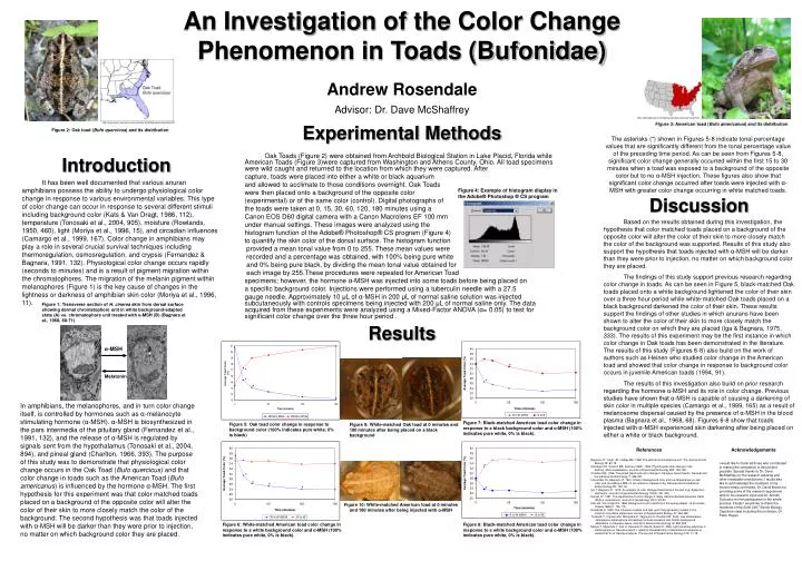
* * * * * * * * Oak Toads (Figure 2) were obtained from Archbold Biological Station in Lake Placid, Florida while American Toads (Figure 3)were captured from Washington and Athens County, Ohio. All toad specimens were wild caught and returned to the location from which they were captured. After capture, toads were placed into either a white or black aquarium and allowed to acclimate to those conditions overnight. Oak Toads were then placed onto a background of the opposite color (experimental) or of the same color (control). Digital photographs of the toads were taken at 0, 15, 30, 60, 120, 180 minutes using a Canon EOS D60 digital camera with a Canon Macrolens EF 100 mm under manual settings. These images were analyzed using the histogram function of the Adobe® Photoshop® CS program (Figure 4) to quantify the skin color of the dorsal surface. The histogram function provided a mean tonal value from 0 to 255. These mean values were recorded and a percentage was obtained, with 100% being pure white and 0% being pure black, by dividing the mean tonal value obtained for each image by 255.These procedures were repeated for American Toad specimens; however, the hormone α-MSH was injected into some toads before being placed on a specific background color. Injections were performed using a tuberculin needle with a 27.5 gauge needle. Approximately 10 μL of α-MSH in 200 μL of normal saline solution was injected subcutaneously with controls specimens being injected with 200 μL of normal saline only. The data acquired from these experiments were analyzed using a Mixed-Factor ANOVA (α= 0.05) to test for significant color change over the three hour period . Figure 4: Example of histogram display in the Adobe® Photoshop ® CS program α-MSH Melatonin * * * Figure 5: Oak toad color change in response to background color (100% indicates pure white, 0% is black) Figure 7: Black-matched American toad color change in response to a black background color and α-MSH (100% indicates pure white, 0% is black). Figure 6: White-matched American toad color change in response to a white background color and α-MSH (100% indicates pure white, 0% is black). Figure 8: Black-matched American toad color change in response to a white background color and α-MSH (100% indicates pure white, 0% is black). An Investigation of the Color Change Phenomenon in Toads (Bufonidae) Advisor: Dr. Dave McShaffrey http://wwknapp.home.mindspring.com/docs/american.toad.html http://www.npwrc.usgs.gov/resource/herps/amphibid/species/oaktoad.htm Figure 3: American toad (Bufo americanus) and its distribution Experimental Methods Figure 2: Oak toad (Bufo quercicus) and its distribution The asterisks (*) shown in Figures 5-8 indicate tonal percentage values that are significantly different from the tonal percentage value of the preceding time period. As can be seen from Figures 5-8, significant color change generally occurred within the first 15 to 30 minutes when a toad was exposed to a background of the opposite color but to no α-MSH injection. These figures also show that significant color change occurred after toads were injected with α-MSH with greater color change occurring in white matched toads. Introduction It has been well documented that various anuran amphibians possess the ability to undergo physiological color change in response to various environmental variables. This type of color change can occur in response to several different stimuli including background color (Kats & Van Dragt, 1986, 112), temperature (Tonosaki et al., 2004, 905), moisture (Rowlands, 1950, 460), light (Moriya et al., 1996, 15), and circadian influences (Camargo et al., 1999, 167). Color change in amphibians may play a role in several crucial survival techniques including thermoregulation, osmosregulation, and crypsis (Fernandez & Bagnara, 1991, 132). Physiological color change occurs rapidly (seconds to minutes) and is a result of pigment migration within the chromatophores. The migration of the melanin pigment within melanophores (Figure 1) is the key cause of changes in the lightness or darkness of amphibian skin color (Moriya et al., 1996, 11). Discussion Based on the results obtained during this investigation, the hypothesis that color matched toads placed on a background of the opposite color will alter the color of their skin to more closely match the color of the background was supported. Results of this study also support the hypothesis that toads injected with α-MSH will be darker than they were prior to injection, no matter on which background color they are placed. The findings of this study support previous research regarding color change in toads. As can be seen in Figure 5, black-matched Oak toads placed onto a white background lightened the color of their skin over a three hour period while white-matched Oak toads placed on a black background darkened the color of their skin. These results support the findings of other studies in which anurans have been shown to alter the color of their skin to more closely match the background color on which they are placed (Iga & Bagnara, 1975, 333). The results of this experiment may be the first instance in which color change in Oak toads has been demonstrated in the literature. The results of this study (Figures 6-8) also build on the work of authors such as Heinen who studied color change in the American toad and showed that color change in response to background color occurs in juvenile American toads (1994, 91). The results of this investigation also build on prior research regarding the hormone α-MSH and its role in color change. Previous studies have shown that α-MSH is capable of causing a darkening of skin color in multiple species (Camargo et al., 1999, 165) as a result of melanosome dispersal caused by the presence of α-MSH in the blood plasma (Bagnara et al., 1968, 68). Figures 6-8 show that toads injected with α-MSH experienced skin darkening after being placed on either a white or black background. Andrew Rosendale Figure 1: Transverse section of H. cinerea skin from dorsal surface showing dermal chromatophore unit in white background-adapted state (A) vs. chromatophore unit treated with α-MSH (B) (Bagnara et al., 1968, 68-71) Results In amphibians, the melanophores, and in turn color change itself, is controlled by hormones such as α-melanocyte stimulating hormone (α-MSH). α-MSH is biosynthesized in the pars intermedia of the pituitary gland (Fernandez et al., 1991, 132), and the release of α-MSH is regulated by signals sent from the hypothalamus (Tonosaki et al., 2004, 894), and pineal gland (Charlton, 1966, 393). The purpose of this study was to demonstrate that physiological color change occurs in the Oak Toad (Bufo quercicus) and that color change in toads such as the American Toad (Bufo americanus) is influenced by the hormone α-MSH. The first hypothesis for this experiment was that color matched toads placed on a background of the opposite color will alter the color of their skin to more closely match the color of the background. The second hypothesis was that toads injected with α-MSH will be darker than they were prior to injection, no matter on which background color they are placed. Figure 9: White-matched Oak toad at 0 minutes and 180 minutes after being placed on a black background References Acknowledgements Bagnara JT, Taylor JD, Hadley ME. 1968. The dermal chromatophore unit. The Journal of Cell Biology 38: 67-79. Camargo CR, Visconti MA, Castrucci AML. 1999. Physiological color change in the bullfrog, Rana catesbeiana. Journal of Experimental Zoology 283: 160-169. Charlton HM. 1966. The pineal gland and color change in Xenopus laevis Daudin. General and Comparative Endocrinology 7: 384-397. Fernandez PJ, Bagnara JT. 1991. Effect of background color and low temperature on skin color and circulating α-MSH in two species of leopard frog. General and Comparative Endocrinology 83: 132-141. Iga T, Bagnara JT. 1975. An analysis of color change phenomena in the leaf frog, Agalychnis dacnicolor. Journal of Experimental Zoology 192(3): 331-342. Heinen JT. 1991. The significance of color change in newly metamorphosed American toads (Bufo a. americanus). Journal of Herpetology 28(1): 87-93. Kats LB, Van Dragt RG. 1986. Background color-matching in the spring peeper, Hyla crucifer. Copeia 1986(1): 109-115. Rowlands A. 1950. The influence of water and light upon the pigmentary system in the common frog Rana temporaria. Journal of Experimental Biology 27: 446-460. Tonasaki Y, Cruijsen JM, Nishiyama K, Yaginuma H, Roubos EW. 2004. Low temperature stimulates α-melanophore-stimulating hormone secretion and inhibits background adaptation in Xenopus laevis. Journal of Neuroendocrinology 16: 894-905. Moriya T, Miyashita Y, Arai JI, Kusunoki S, Abe M, Asami K. 1996. Light-sensitive response in melanophores of Xenopus laevis: I. spectral characteristics of melanophore response in isolated tail fin of Xenopus tadpole. The Journal of Experimental Zoology 276: 11-18. I would like to thank all those who contributed to making the completion of this project possible. Special thanks to Dr. Dave McShaffrey for his research advising and other invaluable contributions. I would also like to acknowledge the members of my honors thesis committee, Dr. David Brown for providing some of the research specimens and for his research input and Dr. Almuth Tschunko for her participation in the whole process. Finally I would like to thank the members of the 2006-2007 Senior Biology Capstone class including the professor, Dr. Peter Hogan. Figure 10: White-matched American toad at 0 minutes and 180 minutes after being injected with α-MSH