Download

1 / 61
650 likes | 1.67k Vues
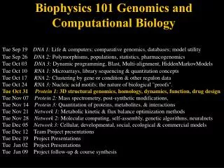
Functional Genomics-Proteomics (I): Overview & Strategies for protein separation. Yao-Te Huang Aug 6, 2010. The website for downloading lecture notes/lecture materials. http://mail.cmu.edu.tw/~ythuang/teaching.summer.2010.htm. Proteome.

E N D
Functional Genomics-Proteomics (I): Overview & Strategies for protein separation Yao-Te Huang Aug 6, 2010
The website for downloading lecture notes/lecture materials • http://mail.cmu.edu.tw/~ythuang/teaching.summer.2010.htm
Proteome • Proteome is a complete set of proteinsproduced by a given cell or organism under a defined set of conditions. • Proteome is a complex and dynamic entity that can be defined in terms of the sequence, structure, abundance, localization, modification, interaction , and biochemical function of its components, providing a rich and varied source of data.
Proteomics • It is the study of proteome. • It is concerned with the systematic, large-scale analysis of proteins. • This analysis requires an equally diverse range of technologies.
The need for proteomics • Nucleic acids are only information-carriers. Thus, the analysis of genes or of mRNA can only tell us about protein function indirectly. • A true understanding of such systems can only come from the direct study of proteins.
The scope of proteomics • Proteins can be studied in various contexts, including sequence, structure, interactions, expression, localization, and modification. • Proteomics is divided into several major but overlapping branches, that embrace these different contexts: (a) sequence & structural proteomics, (b) expression proteomics, (c) interaction proteomics, and (d) functional proteomics.
Requirements of techniques that are suitable for proteomics • High resolution: ruling out one-dimensional techniques • High throughput: (if possible, to resolve all the proteins in one experiment and to be easy to automate) • Be compatible with downstream analysis of mass spectrometry
Two strategies for protein separation at the proteomic level • Two-Dimensional Gel Electrophoresis (2DGE) • Multi-Dimensional Liquid Chromatography (MDLC)
2-dimensional gel electrophoresis permits high resolution of a complex mixture of proteins The first dimension: IEF The second dimension: SDS-PAGE
Proteins from E. coli were separated by two-dimensional gel electrophoresis, resolving more than a thousand different proteins
Isoelectric focusing • Electrophoresis is carried out in a pH gradient, allowing each protein to migrate to its isoelectric point, i.e., the point at which its pI value is equivalent to the surrounding pH and its net charge is zero. • Diffusion still acts against this tendency to focus at a single position in the gel, but a protein diffusing away from its isoelectric point becomes charged and therefore moves back to its focus
Isoelectric focusing • Running the gel for a suitably long period of time ensures that all proteins reach their isoelectric points and size-independent separation is achieved.
The pH gradient in an IEF gel can be established in two ways • To use synthetic carrier ampholytes which are collections of small amphoteric molecules with pI values corresponding to a given pH range. • To use immobilized pH gradient (IPG) strip gels, in which the buffering groups are attached to the polyacrylamide matrix of the gel.
To use synthetic carrier ampholytes • Initially, all the ampholytes are evenly distributed, so there is no pH gradient. When the electric field is applied, however, the ampholytes themselves are subject to electrophoresis. The most acidic ampholyte moves towards the anode (positive electrode), the most basic ampholyte moves towards the cathode (negative electrode) and all the other ampholytes establish intermediate zones according to their pI values. • The proteins can be added to the gel before the electric field is applied or after a period of prefocusing.
Problems associated with the use of synthetic carrier ampholytes • Cathodic drift: the ampholytes themselves migrate to the cathode due to a phenomenon called electro-osmotic flow (bulk solvent movement towards the cathode). This results in pH gradient instability as basic ampholytes are progressively lost from the system. • Poor reproducibility: Because the gel is not run for long time enough to allow the system to reach equilibrium, the proteins are separated in a rapidly forming pH gradient that never becomes stable. For this reason, the conditions of separation are very hard to reproduce.
To use immobilized pH gradient (IPG) strip gels • The IPG is established using immobilines, a collection of nonamphoteric molecules that contain a weak acid-or base-buffering group at one end, and an acrylic double bond to facilitate the immobilization reaction at the other. • These chemicals are available from Amersham Pharmacia Biotech. The gel is run in the normal way but the pH gradient exists before the electric field is applied, and remains stable even when the gel is run for a long time.
SDS-PAGE • All protein-SDS complexes have the essentially the same charge density. • The relative differences in mass between proteins are maintained in the protein-SDS complexes.
Limitations of 2DGE in proteomics • The basic procedure for 2DGE has changed little since 1975 (Patrick O’Farrell), although the rather cumbersome tube gels have been largely replaced by IPG strip gels. • These limitations fall into four major areas: resolution, sensitivity, representation, and automation.
Improving the resolution of 2DGE • Today’s standard 2DGE systems are able to resolve about 2500 protein spots on a routine basis.
Improving the resolution of 2DGE • (1) The resolution of 2DGE depends on the separation length in both dimensions, and can thus be increased if very large format gels are used. For example, 2DGE allows the separation of up to 10000 protein spots if IPG strips >30 cm in length & SDS gels > 30 cm are used.
Improving the resolution of 2DGE • (2a) to use multiple IEF gels, each with a narrow pH range. These are known as zoom gels. IPG 3-12 IPG 5-6
Improving the resolution of 2DGE • (2b) to use the gels with nonlinear pH gradients
Improving the resolution of 2DGE • (3) use various forms of pre-fractionation prior to electrophoresis, to simply the protein mixture that is being analyzed. These may include one or more rounds of chromatography, sucrose density gradient centrifugation, or affinity-based enrichment or depletion procedures.
Improving the sensitivity of 2DGE • The sensitivity problems of 2DGE fall into two categories: • The first is the difficulty in detecting the rarest proteins at all, which reflects the sensitivity of protein staining, detection and quantitation methods.
Improving the sensitivity of 2DGE • The second problem is the tendency of the spots produced by abundant proteins to mask or obscure those produced by scare proteins. • The use of narrow-range IPG gels in combination with pre-fractionation or affinity-depletion of very abundant proteins goes a long way to resolving the problems caused by masking, particularly because this allows larger amounts of the sample to be loaded.
The representation of proteins on 2D-gels • Membrane proteins are under-represented on standard 2D-gels. Possible solutions: using strong detergent (e.g., CHAPS), enriching fractions containing membrane proteins, etc. • Histones, other chromatin proteins, and ribosomal proteins are very hard to separate by standard 2DGE.
The representation of proteins on 2D-gels • A modified 2DGE approach that has been widely used for the separation of histones involves a first separation carried out on an acid-urea gel (which separates the proteins on the basis of size) and the second-dimension separation carried out on an acid-urea-Triton gel. Histones with different hydrophobicity binds differentially to Triton. • Modified buffers are used to break the aggreation of nuclear proteins in 2DGE.
The automation of 2DGE • Capturing the images from stained 2D-gels and then isolating particular spots for further processing and mass spectrometry used to be hard to automate and therefore constitutes the most significant bottleneck in proteomic research.
The automation of 2DGE • There are now various software packages available that produce high-quality digitalized gel images and incorporate methods to evaluate quantitative differences between spots on different gels. • Spot excision robots use plastic or steel picking tips to transfer gel slices to micro-titer plates for automated digestion, clean-up, concentration, and transfer to the mass spectrometer.
The conventional way of doing a typical proteomic experiment.
The requirement for sample separation before running MS: due to sample complexity • (1) 1D-gel electrophoresis (or more commonly 2-D gel electrophoresis) followed by mass spectrometry • (2) 1D LC (or 2D-LC, or MDLC, standing for multi-dimensional liquid chromatography) followed by mass spectrometry
The drawbacks of 2-D gel electrophoresis • being labor-intensive, • selecting against very large and very small, extremely acidic or basic or membrane proteins, • biasing toward highly abundant protein Thus, proteins of medium to low abundance are difficult to detect in 2-D gels. These are typically the proteins, including transcription factors and protein kinases for example, that matter. • Quantitation by 2-D gel method may not be so sensitive or reliable!
Introduction to MDLC • MDLC stands for Multidimensional Liquid Chromatography. • MDLC is distinguished from conventional 1-D chromatographic procedures by comprising a specific set of carefully chosen and matched chromatographic separation modes capable of yielding highly purified proteins in a minimum number of steps. Automation is frequently used to join the various stages of separation into a single efficient procedure.
Strategies of MDLC • Two separate approaches can be defined for MDLC: off-line (discontinuous) and on-line (continuous). Each has its own advantages and disadvantages • For the purpose of discussion, MDLC separations are described as two-dimensional, but they could be extended to more dimensions of selectivity.
Offline-coupling • In offline-coupling, fractions from a first chromatographic separation are collected, either manually or with an automated fraction collector, and then injected into the second separation system. • Off-line coupling is similar to the conventional 1-D protein purification methods, in which multiple stages are used to progressively purify and enrich the sample.
Offline-coupling (contd.) • One advantage of the off-line approach is that the different separation dimensions can be operated independently.
On-line coupling • In on-line coupling MDLC, fractions from the first dimension are transferred directly to the second dimension. • Although this approach requires more complex instrumentation, it has a number of advantages in terms of reproducibility, ease of use, and automation.
On-line coupling (contd.) • A key advantage is that the sample is kept in a closed system under controlled conditions, which can result in improved protein recovery and stability. • A typical on-line coupling MDLC procedure is accomplished using high-pressure multi-position multi-port valves.
On-line coupling (contd.) • The appropriate fractions from the first dimension is transferred to the second dimension, either directly or through one or more intermediate trapping columns. • Five typical valving configurations are shown below:
Possible configurations for 2D-HPLC Case a: Integrated hybrid column Case b: direct serial connection of two separate columns Case c: valved connection with one two-way six-port valve allowing bypass of first dimension
Possible configurations for 2D-HPLC (contd.) Case d: valved connection with two two-way six-port valves allowing isolation of first dimension and selected isolation of fraction on trapping column (T). Case e: valved connection with one two-way six-port valve for selecting or isolating first dimension and two multi-way multi-port valves for selecting or isolating trapping columns 1-8.
In one approach, a sample is initially injected and completely retained on the first dimension. It is then eluted, in discrete fractions, directly onto the second dimension. While the initial eluate is being separated in the second dimension, the rest of the sample remains in the first dimension until it, in turn, is eluted in a stepwise manner and separated in the second dimension. For this approach, the first dimension must be retentive. • If the two dimensions are coupled directly (without a multi-port valve), then the first dimension must remain retentive under all elution conditions of the second dimension. This is because the mobile phase for both dimensions flows continuously through both stationary phases