Download
1 / 32
320 likes | 906 Vues
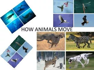
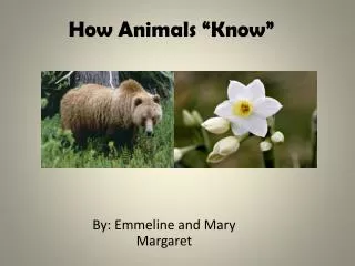
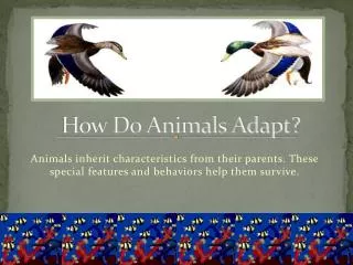
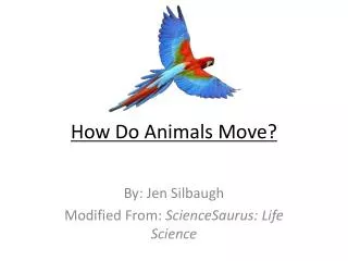
How Animals Move. Chapter 32. Skeleton. A medium or structural element against which contractile cells can act Three types Hydrostatic Exoskeleton Endoskeleton. exoskeleton. Endoskeleton. All vertebrates have endoskeletons Fins or limbs attach to skeleton at pectoral and pelvic girdles.

E N D
How Animals Move Chapter 32
Skeleton • A medium or structural element against which contractile cells can act • Three types • Hydrostatic • Exoskeleton • Endoskeleton exoskeleton
Endoskeleton • All vertebrates have endoskeletons • Fins or limbs attach to skeleton at pectoral and pelvic girdles
s clavicle scapula sternum humerus rib radius vertebral column ulna pelvic girdle femur patella tibia fibula Human Skeleton
Functions of Bone • Interact with muscle to enable movement • Support and anchor muscles • Enclose and protect internal organs • Store calcium and phosphorus • Produce blood cells
Long Bone Structure • Compact bone • Spongy bone • Central cavity contains yellow marrow
Compact Bone Structure • Mature compact bone consists of many cylindrical Haversian systems
Bone Marrow • Yellow marrow • Fills the cavities of adult long bones • Is largely fat • Red marrow • Occurs in spongy bone of some bones • Produces blood cells
Bone Remodeling • In adults, bone building and bone breakdown continue constantly • Osteoblasts deposit bone • Osteoclasts secrete enzymes that degrade it • Remodeling adjusts bone strength and helps maintain blood calcium levels
Bone Density • Exercise can increase bone density • Osteoporosis is a decrease in bone density • May occur when the action of osteoclasts outpaces that of osteoblasts • May also occur as a result of inability to absorb calcium
Joints • Areas of contact or near contact between bones • Fibrous joints • Short connecting fibers join bones • Synovial joints • Move freely; ligaments connect bones • Cartilaginous joints • Straps of cartilage allow slight movement
Skeletal Muscle • Bundles of striped muscle cells • Attaches to bone • Often work in opposition biceps triceps
Tendons Attach Muscle to Bone muscle tendon bursae synovial cavity
TRICEPS BRACHII Human Skeletal Muscles BICEPS BRACHII PECTORALIS MAJOR DELTOID TRAPEZIUS SERRATUS ANTERIOR EXTERNAL OBLIQUE LATISSIMUS DORSI RECTUS ABDOMINUS GLUTEUS MAXIMUS ADDUCTOR LONGUS BICEPS FEMORIS SARTORIUS QUADRICEPS FEMORIS GASTROCNEMIUS TIBIALIS ANTERIOR
Skeletal Muscle Structure • A muscle is made up of muscle cells • A muscle fiber is a single muscle cell • Each fiber contains many myofibrils myofibril
Sarcomere A myofibril is made up of thick and thin filaments arranged in sarcomeres sarcomere sarcomere sarcomere sarcomere Z band Z band Z band
Muscle Microfilaments Thin filaments • Like two strands of pearls twisted together • Pearls are actin • Other proteins in grooves in filament Thick filaments • Composed of myosin • Each myosin molecule has tail and a double head
Sliding-Filament Model Sarcomere shortens because the actin filaments are pulled inward, toward the sarcomere center
Sliding-Filament Model • Myosin heads attach to actin filaments • Myosin heads tilt toward the sarcomere center, pulling actin with them
Role of Calcium in Contraction • T tubules in the sarcoplasmic reticulum relay signal • Calcium ions are released
Nervous System Controls Contraction • Signals from nervous system travel along spinal cord, down a motor neuron • Endings of motor neuron synapse on a muscle cell at a neuromuscular junction
Contraction Requires Energy • Muscle cells require huge amounts of ATP energy to power contraction • The cells have only a very small store of ATP • Three pathways supply ATP to power muscle contraction
Motor Unit • One neuron and all the muscle cells that form junctions with its endings • When a motor neuron is stimulated, all the muscle cells it supplies are activated to contract simultaneously • Each muscle consists of many motor units
Muscle Tension • Mechanical force a contracting muscle exerts on an object • For a muscle to shorten, muscle tension must exceed the load that opposes it • The load may be the weight of an object or gravity’s pull on the muscle
Two Main Types of Contraction • Isotonic contraction • Muscle visibly shortens; moves a load • Tension remains constant as the muscle changes length • Isometric contraction • Muscle does not change length • Tension is insufficient to move load
Muscle Fatigue • An inability to maintain muscle tension • Occurs after a period of tetanic contraction • Different types of muscle show different fatigue patterns
Muscular Dystrophies • A class of genetic disorders where muscles progressively weaken and degenerate • Duchenne muscular dystrophy is the most common among children • Myotonic muscular dystrophy is the most common among adults
Aging Muscles • Over time, the number and size of muscle fibers decreases
Clostridium • Clostridium botulinum – causes botulism, stopping the release of acetylcholine, a neurotransmitter that enables muscle contraction • Clostridium tetani – causes tetanus, blocking the neurotransmitters GABA and glycine, which leads to uninhibited muscle contraction