Download
1 / 48

E N D
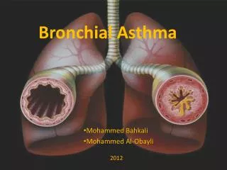
BRONCHIAL ASTHMA Prof. Vatutin N.T.
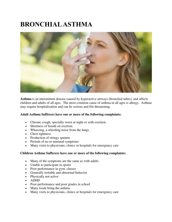
Definition Asthma is a chronic inflammatory disease of the airways which develops under the allergens influence, associates with bronchial hyperresponsiveness and reversible obstruction and manifests with attacks of dyspnea, breathlessness, cough, wheezing, chest tightness and sibilant rales more expressed at breathing-out.
Epidemiology According to epidemiological studies asthma affects 1-18% of population of different countries. Only in 2006 more than 300 million patients suffered from asthma all over the world, 250 thousands of patients die of asthma. The incidence of asthma is higher in countries with increased air pollution.
As asthma is a respiratory allergic disease, the influence of allergens permeated into the organism through airways is essential for the disease development. The allergens are divided into: Etiology • communal, • industrial, • occupational, • natural • pharmacological
Сommunal allergens are contained in the air of apartment houses. They are: • house-dust mites which live in carpets, mattresses and upholstered furniture; • spittle, excrements, desquamated epidermis, hair and fur of domestic animals; • vital products of domestic insects (e.g., cockroach); • mycelial yeast-like fungi (molds); • tobacco smoke during active or passive smoking; • various communal aerosols and synthetic detergents.
Among the industrial allergens nitric, carbonic, sulfuric oxides, formaldehyde, ozone and emissions of biotechnological industry - main components of industrial and photochemical smog - must be mentioned. The most important occupational allergens are dust of stock buildings, mills, weaving-mills, book depositories etc. Natural allergens are represented by plant pollen (especially ambrosia, wormwood and goose-foot pollen) and different respiratory, particularly viral, infections.
Some allergens which may cause asthma Spittle, excrements, hair and fur of domestic animals House-dust mites which live in carpets, mattresses and upholstered furniture Plant pollen Dust of book depo-sitories Food components (stabilizers, genetically modified products) Pharmacological agents (enzymes, antibiotics, vaccines, serums)
Genetic abnormalities which lead to excessive production of allergen-specific antibodies (Ig E) is called atopy - significant factor in asthma genesis in many patients. A lot of cases of atopy are inherited, and recent epidemiological studies showed steady increase in number of people with high serum level of Ig E. Trigger-factors, which provoke bronchospasm, are: a simultaneous penetration of a large quantity of allergen, viral respiratory infection, hyperventilation, physical exertion, emotional stress, becoming too cold, adverse weather conditions, administration of some medicines (aspirin, b-blockers).
Pathophysiology Asthma pathophysiology is quite difficult and insufficiently studied. Undoubtedly, in most cases the disease is based on 1 type hypersensitivity reaction. The genesis of any allergic reaction may be divided into immune, pathochemical and pathophysio-logic phases.
Allergens Immune phase Allergen-specific IgE B-cell After involving into the airways allergens activate immunocompetent cells. As a result B-lymphocytes produce antibodies of Ig E class. In case of asthma T-lymphocytes are inhibited, so the activation of B-lympocytes and Ig E production are excessive, exceeding normal needs. T-cell
Further these antibodies bind to the surface of mast cells, basophils and eosinophils of bronchial mucous. When a new portion of allergen involves the respiratory system, it interacts with IgE-antibodies. This is a first, immune phase of allergic reaction.
Pathochemical phase As a result of antigen-antibody reaction the peculiar “explosion” occurs. The membranes of mast cells, basophils and eosinophils of bronchial mucous wreck with output of biologically active substances (histamine, serotonin, chemotaxis factors, heparin, proteases, thromboxane, leukotrienes, prostaglandins), which induce hyperergic inflammation, mucous edema, spasm of smooth myocytes, glands hypersecretion, viscous exudate formation in bronchial lumen. Airway fill with mucus Muscles contract Airways swell
Asthma exacerbation, occurring as a result, is a clinical manifestation of the 3rd, pathophysiolo-gical, phase of allergic reaction. The indicated mechanism is specific for atopic (exogenous) asthma genesis. In addition to this,autosensibilization of damaged pulmonary tissue, neuropsychic disturbances, corticoid insufficiency, adrenergic imbalance, impairment of arachidonic acid metabolism, genetic and some other factors probably play a certain role in genesis of nonatopic (endogenous) asthma.
Pathologic anatomy Macroscopic changes: • viscous mucous/ mucopurulent phlegm • airway dyskinesia with zones of spastic contraction and paralytic expansion of bronchi • obstruction of airway lumen • lung emphysema, pneumosclerosis • RVand RA hypertrophy and dilation
Microscopic changes: • Bronchial wall infiltration with mast cells, eosinophils, basophils and T-lymphocytes • Edema of mucous and submucous tunics • Destruction of bronchial epithelium • Hypertrophy of bronchial smooth muscles, • Hyperplasy of submucous glands • Microvessels dilation
Classification Depending on etiology asthma is divided into exogenous (atopic) and endogenous (non-atopic).Byclinical course asthma is divided into intermittent (beginning, early) and persistent (chronic, late). Depending on frequency of exacerbations, limitations of patient’s physical activity and lung function persistent asthma is divided into mild, moderate and severe (lung function is assessed by forced expiratory volume in 1 second (FEV1) and peak expiratory flow (PEF) and daily variability of these parameters). There are also remission phase and exacerbations.
Clinical course, severity Daytime asthma symptoms Nighttime awakenings FEV1, PEF Intermittent < 1 /week 2 and < /month >80% predicted. Daily variability < 20% Mild persistent 1 /week but not daily > 2 /month >80% predicted. Daily variability – 20-30% Moderate persistent Daily > 1 /week > 60 but < 80% predicted. Variability>30%. Severe persistent Persistent, which limit normal activity Daily <60% predicted. Variability > 30%. Asthma severity classification
In recommendations of Global Initiative for Asthma (GINA) asthma is classified on the base of control assessment andis divided into well-controlled, partially controlled and uncontrolled. Asthma control is considered as: • daytime symptoms 2 /week; • ability to engage in normal daily activity; • the absence of nighttime awakenings as a result of asthma symptoms; • need in bronchodilators administration 2 /week; • the absence of asthma exacerbations; • normal or near normal lung function parameters.
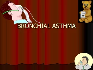
Clinical manifestations Classic signs and symptoms of asthma are: • attacks of expiratory dyspnea • shortness of breath • cough • chest tightness • wheezing (high-pitched whistling sounds when breathing out) • sibilant rales
In typical cases in development of asthma exacerbation there are 3 periods – prodromal period, the height period and the period of reverse changes. At theprodromal period: • vasomotoric nasal reaction with profuse watery discharge, • sneezing, dryness in nasopharynx, • paroxysmal cough with viscous sputum, • emotional lability, • excessive sweating, • skin itch and other symptoms may occur.
At the peackof exacerbation there are: • expiratory dyspnea • forced position with supporting on arms • poorly productive cough • cyanotic skin and mucous tunics • hyperexpansion of thorax with use of all accessory muscles during breathing • at lung percussion: tympanitis, shifted downward lung borders • at auscultation: diminished breath sounds, sibilant rales, prolonged breathing-out, tachycardia. • in severe exacerbations: the signs of right-sided heart failure (swollen neck veins, hepatomegalia), overload of right heart chambers on ECG.
At the period of the reverse changes, • which comes spontaneously or under pharmacologic therapy, • dyspnea and breathlessness relieve or disappear, • sputum becomes not so viscous, • cough turns to be productive, • patient breathes easier.
Asthmatic status The severe and prolonged asthma exacerbation with intensive progressive respiratory failure, hypoxemia, hypercapnia, respiratory acidosis, increased blood viscosity and the most important sign is blockade of bronchial b2-receptors. Stages:1st- refractory response to b2-agonists (may be paradoxical reaction with bronchospasm aggravation) 2nd - “silent” lung because of severe bronchial obstruction and collapse of small and intermediate bronchi; 3rd stage – the hypercapnic coma.
In many cases asthma, particularly intermittent, manifests with few and atypical signs: • episodic appearance of wheezing; • cough, heavy breathing occurring at night; • cough, hoarseness after physical activity; • “seasonal” cough, wheezing, chest tightness (e.g., during pollen period of ambrosia); • the same symptoms occurring during contact with allergens, irritants; • lingering course of acute respiratory infections.
The complications of asthma exacerbations are: pneumothorax lung atelectasis pneumonia acute or subacute cor pulmonale asthmatic status. Persistent asthma causes: fibrosing bronchitis small bronchi deformation and obliteration emphysema pneumosclerosis, chronic respiratory failure chronic cor pulmonale. Asthma complications Asthma in childhood leads to growth inhibition and thoracic deformation.
Investigations Lab Data Eosinophilia, moderate leukocytosis in blood count as well as increased serum level of Ig E can be found in patients with asthma, especially at asthma exacerbations. Inflammatory cells, Curschmann's spirals (viscous mucus which copies small bronchi) and Charcot-Leyden crystals (crystallized enzymes of eosinophils and mast cells) can be observed in sputum.
Chest X-ray reveals: • hyperlucency of lung fields • low standing and limited mobility of diaphragm • expanded intercostal spaces • horizontal rib position.
ECG especially in case of severe, persistent asthma, shows hypertrophy of right heart chambers. Right axis deviation, Rs type complex in V1 lead, low amplitude R in V5-V6 leads
Lung function assessment The diagnosis and severity assessment of asthma is based mainly on parameters of lung function.The most important of them are: forced expiratory volume in 1 second (FEV1) and peak expiratory flow (PEF), which • are measured during spirometry at forced breathing-out.
PEF FEF FEV1 and PEF directly depend on bronchial lumen size and elastic properties of surrounding lung tissue. Expiration FEF Flow Volume Inspiration PIF
Increase in FEV1 and PEF after inhalation of bronchodilators (b2-agonists) of 15% and more is specific for asthma.
PEF also can be measured with the help of individual devices – peak flow meters
Diagnosis Typical clinical manifestations and lung function assessment are sufficient for diagnosis of asthma.
Differential diagnosis Asthma is to be differentiated with a number of diseases manifesting with dyspnea – cardiac, uremic, hysteric asthma, systemic vasculitis, broncho-carcinoma, carcinoid and chronic obstructive lung diseases.
In contrast to bronchial, in case of cardiac asthma the signs of severe heart disease and pulmonary congestion can be found.
In case of uremic asthma severe renal pathology takes place as well as other manifestations of uremia. Dyspnea without bronchospasm is specific for hysteric asthma. In patients with systemic vasculitis fever, weight loss, impairment of other organs and nervous system, arterial hypertension, arthralgia, myalgia, specific changes in tissue sampling may be revealed. Carcinoid intestinal tumors manifest with watery excrements, rumbling, heat sensation in head, tricuspid valve damage. In differentiating of bronchospastic syndrome in bronchial tumors ray-path methods (X-ray, computer tomography, bronchography) and bronchoscopy are importantant.
Management includes: 1. Avoiding the contact with allergen. If it is impossible, the specific hyposensitization with standard allergens should be performed. It is rather effective in case of monoallergy, in intermittent and mild persistent asthma, in remission phase. 2. Elimination of trigger factors (rational job placement, changing the residence, psychological and physical adaptation, careful drug using) is the second condition for successful asthma treatment. 3. Optimally selected medical care is the base of asthma management.
2 drug categories are used: Drug therapy Antiinflammatory drugs (basic) Bronchodilators Are divided into: 3 groups: hormone-containing (corticosteroids) b2-agonists anticholinergic drugs nonhormone-containing (cromones, leukotriene receptor antagonists) methylxanthines
The working mechanism lays in: cell membrane stabilization inhibition of inflammatory mediators restoring the sensivity of b2-receptors. Inhaled corticosteroids (beclamethazone, inhacort, budesonide, flixotid, fluticazone, asmacort, asthmanex) are the most effective and safe and considered to be the first line drugs for asthma treatment. Systemic are used during short courses, mainly in case of severe persistent asthma or asthmatic status. Corticosteroids
Cromones (cromolyn sodium – intal, and nedocromil – tiled) stabilize cell membranes, used mainly in pediatric practice (in childhood) in case of intermittent or mild persistent asthma. Leukotriene receptor antagonists (montelukast, zafirlukast) have the moderate intiinflammatory activity used in case of aspirin-induced asthma and asthma of physical exertion.
Bronchodilators b2-agonists Anticholinergic drugs Stimulates b2-adrenergic receptors of bronchi reduce tonus of vagus Smooth muscle relaxation Methylxanthines inhibit phosphodiesterase
Inhaled b2-agonistsare the basic drug group among bronchodilators. • Short-acting (duration of action 5-6 h) b2-agonists - salbutamol, fenoterol - are used for quick relief of asthma symptoms. • Long-acting (> 12 h) b2-agonists - salmoterol, farmoterol - for prevention of asthma symptoms occurring.
Anticholinergic drugs (ipratropium bromide, atrovent, troventol) are used predominantly in nighttime asthma and in elderly patients because of the least cardiotoxic effect. • Methylxanthinesin comparison with other bronchodilators have the less bronchodilating potential. There are long-acting (>12 h) - (theopec, theolong, theodur, euphilong) as well as short-acting (aminophylline, theophylline) drugs in this group.
Combined inhaled drugs (corticosteroids with b2-agonists) – seretid, simbicort – with use of delivery devices (nebulasers, turbuhalers, spasers, spinhalers, sinchroners) enhance the effectiveness of asthma therapy.
Management of asthmatic status • Oxygen • Systemic corticosteroids (Hydrocortisone 200mg or Methylprednisolone 125mg every 6h IV or Prednisolone 50 mg/day per os) • Inhalations of short-acting b2-agonists - Salbutamol 5mg or Fenoterol 2mg through nebulaser – 3 times at 1st hour, then once an hour till distinct improvement of patient’s condition is achieved; then 3-4 times a day. • Inhaled anticholinergic drugs or Aminophylline IV. • If ineffective - artificial lung ventilation.
Prognosis • In case of early detection and adequate treatment the prognosis for the disease is favourable. • It becomes serious in severe persistent and poorly controlled (insensitive for corticosteroids) asthma.
The examination of working capacity • The patients with unfavorable for the disease conditions of work need the job replacement. • Physical labours with severe asthma are disable to work.
Prophylaxis Preservation of the environment, healthy life-style (smoking cessation, physical training) – are the basis of primary asthma prophylaxis. These measures in combination with adequate drug therapy are effective for secondary prophylaxis.