Download
1 / 53
640 likes | 1.7k Vues
UNDERGRADUATE COURSE LECTURERS IN OBSTETRICS AND GYNECOLOGY ,Faculty of medicine ,Zagazig University

E N D
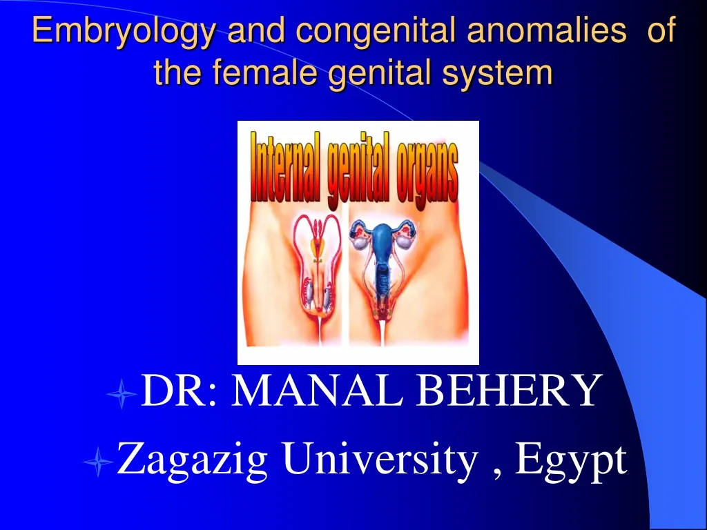
Embryology and congenital anomalies of the female genital system • DR • DR: MANAL BEHERY • Zagazig University , Egypt
C Components which form female and male reproductive systems are: 1. Gonads Ovaries or testes 2. Genital Duct Systems Mesonephric and Paramesonephric ducts 3. External Genitalia
Genotype of embryo 46XX or 46XY is established at fertilization • SRY (sex-determining region on Y) that encodes a protein called testis-determining factor (TDF) are responsible for male differentiation
Indifferent Embryo • Weeks 1-6 sexually indifferent or undifferentiated stage • Week 7 begins phenotypic sexual differentiation • Week 12 female or male characteristics of external genitalia can be recognized and completed at 20 weeks.
At 4TH Week of gestation • Mesonephric Duct • extending from the • mesonephros • (Wolff’s body) • to the cloaca • (urogenital sinus)
A Swelling on either side of dorsal mesentry on medial side of mesonephric duct forms the urogenital ridge At 5TH Week of gestation
Primitive germ cells migrate from yolk sac through dorsal mesentry to reach genital ridge These germ cells stimulate coelomic epithelium and underlying mesoderm to proliferate and form primitive sex cords Formation and Differentiation of Gonads
At 6TH week gestation Paramesonephric or Mullerian Duct develops lateral to the Mesonephric ”wolffian “ Duct
The middle and caudal parts of the Mullerian ducts undergoes medial migration and fusion. • The cranial 1/3 → tubes. • The middle 1/3 → uterus and cervix. • Caudal 1/3 → upper 3/4 of vagina.
2 main Principle • Internal genital organs develop in close association with urinary tract So gross malformation of uterus and tube are commenaly associated with anomalies of kidney and ureter. • 2-Development of gonads is separt from that of the ducts So functional ovary are usually present when uterus, vagina are absent
In ovary the absence of testosterone inhibits the development of the mesonephric ducts. The atretic remains form the epoophoron, paraoophoron and Gartner’s ducts. In absence of AMH, paramesonephric ducts form the female internal genital tract. Female Genital Duct Formation
Abnormalities of the ovaries: • 1) agenesis or complete absence. • 2) Gonadal dysgenesis "streak gonads" as in Turner syndrome. • 3) Failure of descent into the pelvis. • 4) Ovotestis “true hermaphrodite” In which combined ovarian and testicular tissues seen.
Development external genitalia • Early, similar in both sexes • 6th wk, three external protuberance surround cloacal membrane, the left and right genital swellings meet anteriorly to form the genital tubercle. • 12th wk identify difference. • Genital swelling labioscrotal folds scrotum or labia major • Genital tubercle phallus penis or clitoris
Why is this important? • Majority have no problem conceiving, but have higher rates of: • 1. Spontaneous Abortion • 2. Premature Delivery • 3. Infertility • 4. Abnormal Fetal Lie • 5. Dystocia at delivery • 6. Dysmenorrhea, endometriosis • 7. Cervical incompetence
Uterine Anomalies Absence of Uterus Fusion anomalies
Classification into 4 groups: • 1. Agenesis of uterus/vagina: Rokitansky-Kuster-Hauser Syndrome. • 2-Unilateral development :Unicornate uterus • 2. Defects in Vertical Fusion (obstructive or non-obstructive) • 3. Lateral Fusion defects (obstructive or non-obstructive).
RKH Syndrome: Diagnosis • Expected Menarche • Difficult to differentiate from imperforate hymen • No uterus on exam, U/S, MRI, Laparoscopy, IVP • Confused with Androgen Resistance Syndrome with shallow pouch and no uterus. • Determine karyotype.
Lateral Fusion Defects: Most common type of mullrian defects
Lateral Fusion Defects • . • Result from failure of fusion of the mullerian ducts • , or failure of • absorption • of the septum. • .
Septate Uterus • Defective resorpation of the septum between the fused mullerian ducts results in a uterine septum, which may extend either partially down the uterus or the full length to the cervix. Normal external surface
Bicornuate Uterus • Fundus indented • Partial fusion of mullerian ducts • Variable degree of separation of uterine horns that can be complete, partial or minimal • HSG not diagnostic , need laparoscopy • problems, however can have
Vertical Fusion Defects: obstructive and non-obstructive • . Can be considered in two categories: • 1.Imperforate Hymen • 2.Transverse Vaginal Septum
Cyclic pelvic pain due to hematocolpos hematometria, or hematosalpinx Bulging hymeneal membrane or a blind-ending pouch on exam. Pelvic/Rectal exam, U/S, MRI Rarely urologic anomalies. Tx: Cruciate incision Imperforate Hymen: Diagnosis/ Treatment
Transverse Vaginal Septum: Presentation/Diagnosis • Cyclical pain due to hematocolpos or hematometria. • Blind-ending pouch. • No bulging at outlet, hydromucocolpos or hematocolpos, rectal exam or U/S, MRI. • Thickness varies and site varies in vaginal canal.
Summary • Understanding the embryologic origin of the defect of mullerian anomalies is key to its correct diagnosis • Presentation: Obstetrical problems, dysmenorrhea, amennorhea • Diagnosis : Pelvic/Rectal exam, U/S, HSG, Laparoscopy, Hysteroscopy, MRI
❍ Where are Gartner ducts located? ❍ Gartner duct cysts are persistent portions of what embryonic structure? ❍ The portion of the gubernaculum between the ovary and uterus becomes what structure? ❍ The portion of the gubernaculum between the uterus and the labium majus becomes what structure?
❍ Where are Gartner ducts located? In the lateral walls of the vagina. ❍ Gartner duct cysts are persistent portions of what embryonic structure? Mesonephric duct. ❍ The portion of the gubernaculum between the ovary and uterus becomes what structure? The ligament of the ovary (utero-ovarian ligament). ❍ The portion of the gubernaculum between the uterus and the labium majus becomes what structure? The round ligament.
❍ Failure of the development of adhesions between the uterus and what structure can result in the ovary migrating through the inguinal canal to the labium majus? ❍ What is the name of a pouch of peritoneum analogous to the saccus vaginalis in the male, which accompanies the gubernaculum in the inguinal canal?
❍ Name the three coats of the ureter. ❍ The epithelium lining the ureter is of what type?