Download
1 / 5

E N D
1 Bone cells Bone cells A And nd their origin their origin Prepared by: Prepared by: Dr. Mohammed Alruby Dr. Mohammed Alruby ناكرب فلا اهفوج يفو ةتماص ضرلااف نايسن تمصلا نا نظت نا كايا B Bone one cells and their origin cells and their origin Dr. Mohammed Alruby Dr. Mohammed Alruby
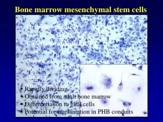
2 Osteoprogenitor cells: pre-osteoblast, are bone stem cells derived from mesenchymal cells that eventually differentiate into mature osteoblast and osteocyte. Osteoblast: large metabolically active cell with increased endoplasmic reticulum(ER) 1-Produce high level of alkaline phosphatase. 2-Produce type I collagen which is necessary for calcification. 3-Produce osteocalcine, produce signal to activate osteoclast. = osteoblast has receptors for hormones such as parathyroid hormone, Vit. D, osteogen, cytokines and growth factors = after osteoblast have secrete un-mineralized bone they usually become inactive, a few osteoblasts remain in the mineralized osteoid and become osteocyte. Osteocyte: are osteoblast that have become surrounded by the calcified matrix of bone, these cells acts as mechanoreceptor identifying the loads placed on the individual bones and establishing the nature of such loads. Osteoclasts: are large multi-nucleated cells, found attached to the surface of active bone formation. = Found in well-defined pits known as Howships Lacuna. = Derived from mono-nuclear stem cells in bone marrow and travel through blood vessels to the site of activity. It is activated by: inter-luckin II,I, cytokines. = decreased endoplasmic reticulum. Bone lining cells: elongated cells covering bone surface, they are inactive and have a high nucleus to cytoplasmic ratio, these cells has a major impact on calcium metabolism within the body. Bone development: Cellular mechanisms: = skeleton formation begins when mesenchymal cells migrate to the site of skeleton-genesis. The cells then interact with epithelial cells, which in then trigger the mesenchymal cells to cluster together and undergo condensation to form compact mass of cells. = each step is regulated by special type of genes such as member of home box genes. = condensed cell then undergo differentiation either chondrocyte or osteoblast. Core bonding factor-1 (CBFA-1)— (now known as Runx2) One of the most important bone specified genes in differentiation of mesenchymal cells into – osteoblast. Core bonding factor -1: CBFA-1 now is known as Runx2. One of the most important bone specific genes in differentiation of mesenchymal cells into------ osteoblast. Bone morphogenetic protein: BMP: = Play important role in the developing skeleton. = BMP has been used ti improving healing and bone defect. = BMP’s are probably involved in intramembranous bone formation. = BMP-7 is found in area of brain to induce formation of cranial bones B Bone one cells and their origin cells and their origin Dr. Mohammed Alruby Dr. Mohammed Alruby
3 = BMP’S 2—4 and 5 are expressed in some regions where mesenchymal condensation later give rise to craniofacial bone. Novel mechanisms of osteoblast and osteoclast interaction: Osteoblast interact with osteoclast to regulate the osteoclastic action. Receptor activator of nuclear factor ligand (RANKL) is produced by pre-osteoblast and osteoblast and cell membrane of osteoblastic precursors. This factor is essential factor for differentiation, fusion into multinucleated cells, activation and survival of osteoclastic cells. Factors regulate skeletal metabolism = Prostaglandin: potent mediator of bone resorption. They can be found in sites of inflammation; it can be demonstrated in: Dentigrous cyst Inflamed gingival tissue Synovial fluid Squamous cell carcinoma Rheumatoid joint = Leukotrienes: action in both bone destruction and bone formation = Cytokines: soluble mediator modulate activity on the same cells. = Inter-leukin -1: IL1: 1- potent stimulator for bone resorption acting both directly or by increasing prostaglandin synthesis 2-Inhibitor of bone formation. = Tumor necrosis factor (TNF) alpha, beta: Stimulate bone resorption and inhibit bone collagen and non-collagenous protein synthesis. It is found in inflammatory lesion like: periodontal disease. = Interleukin 6: found on inflammatory lesion. = parathyroid hormone: released from parathyroid gland in response to low serum calcium low serum phosphate. Low vit D3 = Calcitonin: released from thyroid c-cells in response to high serum Ca, inhibite bone resorption by osteoclast = Growth hormone: important regulator of Ca absorption from intestine and renal tubules. = Testosterone: normal level increase bone growth (in the presence of growth hormone), but high level increase maturation of cells: decrease bone growth. = Estrogen: ovariectomy decrease bone mass—post menopausal female frequently suffer from bone loss which can lead to osteoporosis. Increase level of estrogen, increase maturation of cells and decrease bone resorption of osteoclasts. B Bone one cells and their origin cells and their origin Dr. Mohammed Alruby Dr. Mohammed Alruby
4 Summary of bone disease A-Congenital and hereditary disorders: Cleido-cranial dysplasia: Characterized by skeletal anomalies such as; Late closure of cranial suture Maturation of primary teeth are normal Permanent dentition delayed from 1—4 years Most patients have supernumerary teeth Craniocytosis: Described collection of approximately 70% of congenital disorder that are caused by a premature fusion Aperts: characterized by crniosynostosis of several suture, cleft palate 25% and mental hand cap Achondroplasia: the commonest form of skeletal dysplasia leading to final height 132--- for males and 152—for femals. Osteo-petrosis: rare disorder caused by marked decreased in bone resorption, characterized by lack of osteoblast function. Bone marrow cavities are invaded with bones which frequently causes secondary anemia. Pcnodystosis: rare sclerosing bone disorder characterized by: short status, brachycephaly, open cranial suture, multiple fracture of long bones Osteogenesis imperfect: Type I: childhood type: the most common form of OI and are associated by brittle bone composed of immature woven bone. The number of fracture are variable and heal with good callus formation Sclera are thin and may be appear blue to the visible pigmented coronoids Type II: perinatal type: Lethal with multiple fracture at birth and blue sclera Type III: infant born with features and deformity. Type IV: similar to type I except the sclera is white stature short and dentino genesis imperfect is common Vitamin D3 resistance Rickets: Growth retardation and childhood rickets. B-Metabolic bone disease Osteoporosis: porous bone occurs when the spaces between the trabecular bone become bigger, making it fragile and increased risk of fracture. Treatment: drugs inhibit resorption; bisphosphonate, estrogen, calcitonin Frequency: affect 1 in 3 women. And 1 in 12 men over 50 years in UK Primary hyper parathyrodism; In which one or more parathyroid glands produce too much parathyroid hormone. All patients treated with surgery 50% of patient present with renal stone Osteomalcia and Vit D dependent rickets: Softening and weakening of the bones, caused by lack of vit D, phosphate and calcium B Bone one cells and their origin cells and their origin Dr. Mohammed Alruby Dr. Mohammed Alruby
5 Acromegaly: Greek term means elongated extremities Hypersecretion of growth hormone Nasal bone enlarges, lower jaw protruded, Spacing in teeth Frequency: 55—69 case // million population Cause; most typically by pituitary adenoma. Paget’s disease: Excessive bone turns over in which bone become enlarged and deformed. Features: fracture, neoplasia, nerve compression, high output cardiac failure Frequency: 5 in 100 people over 50 years in UK B Bone one cells and their origin cells and their origin Dr. Mohammed Alruby Dr. Mohammed Alruby