Download
1 / 22
300 likes | 788 Vues
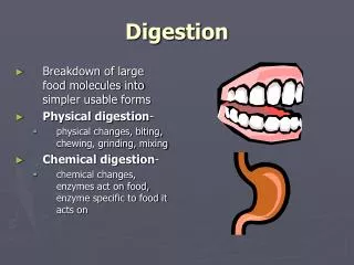
DIGESTION. Digestion : The mechanical and chemical processes taking place in the gastrointestinal tract by which food is broken down into absorbable forms. Digestion is related to, but not the same as metabolism.

E N D
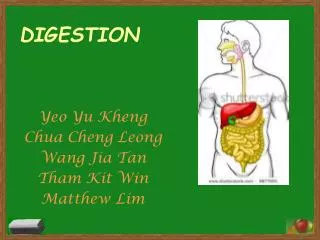
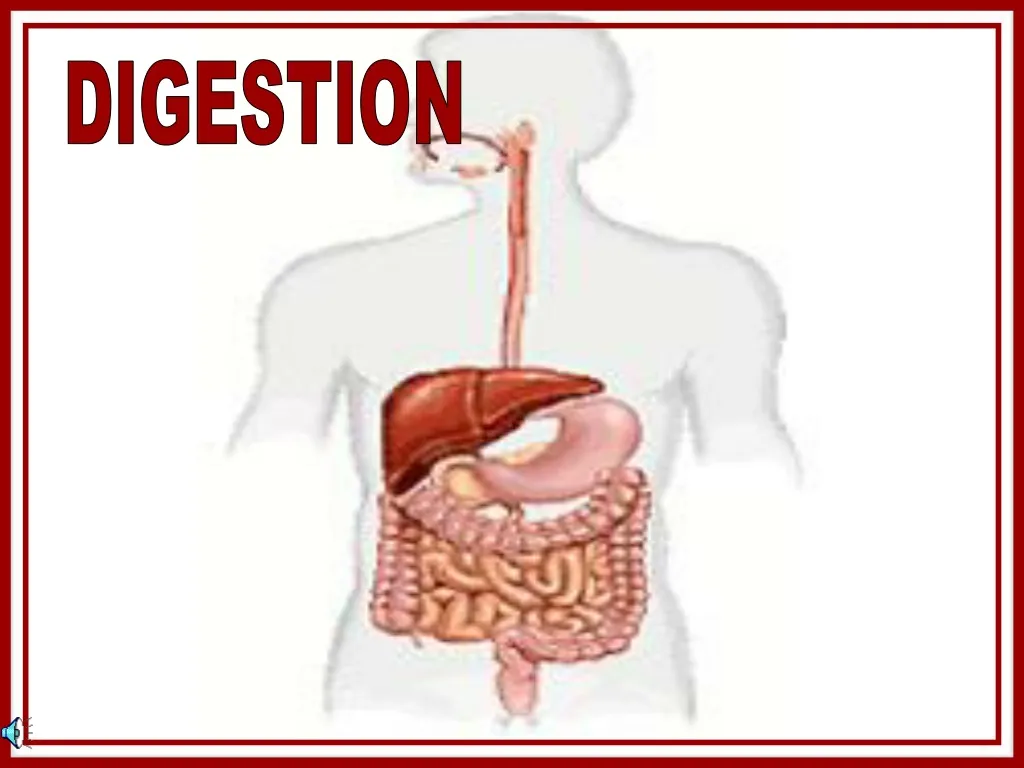
Digestion: The mechanical and chemical processes taking place in the gastrointestinal tract by which food is broken down into absorbable forms. Digestion is related to, but not the same as metabolism. Metabolism: The physical and chemical processes by which substances in food are produced or transformed into energy or products for the uses of the body. The gastrointestinal tract (GI tract), also called the digestive tract, or the alimentary canal, is the system of organs that takes in food, digests it to extract energy and nutrients, and expels the remaining waste. The major functions of the GI tract are digestion and excretion.
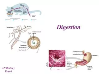
The mouth, also known as the buccal cavity(bŭk'əl)or oral cavity, is the orifice through which we take in food and water. It is the entrance to the digestive tract, and is lined with mucous membranes called buccal mucosa. The membrane-covered roof of the mouth is called the palate. The front part consists of a bony portion called the hard palate, with a fleshy rear part called the soft palate. Mouth... In response to the sensory stimulation of seeing, smelling, tasting, or even imagining good-tasting food, the brain sends impulses through the nerves that control the salivary glands located under the tongue and near the lower jaw, telling them secrete saliva. As the teeth tear and chop the food, saliva moistens it for easy swallowing. A digestive enzyme called amylase (ăm'ə-lāce‘)or ptyalin (tī'ə-lĭn)which is found in saliva, starts to break down some of the carbohydrates (starches and sugars) in the food even before it leaves the mouth.
The tongue is mainly composed of muscles. It is covered with a mucous membrane. Small nodules of tissue called papillae ( pə-pĭl'ē) cover the upper surface of the tongue. Mouth... Between and on the papillae are the taste buds, which provide the sense of taste. In addition to taste, the tongue functions in moving food to aid chewing and swallowing. The human tongue can detect five basic taste components: sweet, sour, salty, bitter, and umami (ū-mä'mē)(savory). The lips and inside of the cheek work with the tongue to position the food for chewing. Mastication or chewing is the repetitive sequence of jaw opening and closingis the process by which food is mashed and crushed by teeth. It is the first step of digestion and it increases the surface area of foods to allow more efficient break down by salivary enzymes and gastric acids. There are several types of teeth, each performing a different function. Incisors cut foods when you bite into them. The sharper and longer canines tear food. The premolars or bicuspids, which are flatter than the canines, grind and mash food. Molars, with their points and grooves, are responsible for the most vigorous chewing.
Pharynx... The pharynx (făr'ĭngks or fair-inks)(plural: pharynges) is the part of the neck and throat situated immediately posterior to the mouth and nasal cavity. Because both food and air pass through the pharynx, a flap of connective tissue called the epiglottis (ĕp'ĭ-glŏt'ĭs)closes over the larynx (lăr'ĭngks or lair-inks)(voice box) opening when food is swallowed. This prevents choking or aspiration, the entry of secretions or foreign material into the trachea (trā'kē-ə)(windpipe) and lungs. Swallowing is a complex act that involves the coordinated activity of the mouth, pharynx, larynx and esophagus. Trachea
The uvula is a small piece of soft tissue that can be seen dangling down from the soft palate over the back of the tongue. It has its own muscle to help it stiffen and change shape, so it fills the back of the throat. . Uvula... It helps keep food from going down the wrong way down the breathing passage when one swallows.
Esophagus... The esophagus, or gullet, is an organ which consists of a muscular tube through which food passes from the pharynx to the stomach. When food is not passing through, the esophagus is folded in, or collapsed.After food is chewed into a mass, a bolus, it is swallowed to move it past the upperesophageal sphincter muscle and into the esophagus. Smooth muscles will contract behind the bolus to prevent it from being squeezed back onto the mouth, then rhythmic, downward-directional waves of contractions called peristalsis will work to rapidly force the food into the stomach. The primary peristaltic wave lasts about 8-9 seconds, even if the bolus descends at a faster rate. In the event that the bolus gets stuck or moves slower than the primary peristaltic wave (as can happen when it is poorly lubricated with saliva), a local reflex response causes a secondary peristaltic wave around the bolus. This secondary wave(s) will force the bolus further down the esophagus, and will continue indefinitely until the bolus passes the lower esophageal sphincter muscle and enters the stomach. (spelled Oesophagus in Greek)
Stomach... Esophagus Fundus The stomach is a bean-shaped hollow muscular organ of the gastrointestinal tract, involved in the second phase of digestion, following mastication. The words gastro- and gastric are derived from Greek, meaning “related to the stomach”. The stomach is usually a highly acidic environment due to gastric acid production and secretion which produces a pH range usually between 1 and 4, depending on food intake, time of the day, drug use, and other factors. Such an environment is able to break down large molecules (such as from food) to smaller ones so that they can eventually be absorbed from the small intestine. The stomach can produce and secrete about 2 to 3 liters of gastric acid per day. Lower Esophageal sphincter Antrum Two smooth muscle valves, or sphincters, keep the contents of the stomach contained. They are the loweresophageal sphincter at the top of the stomach and the pyloric sphincter dividing the stomach from the small intestine. A sticky alkaline mucus is secreted that coats and lubricates the entire gastric surface.
Stomach... The stomach is an organ between the esophagus and the small intestine. It has three tasks. 1. The stomach serves as a short-term reservoir. It stores swallowed food, allowing a large meal to be consumed quickly and dealt with over a period of time. The stomach absorbs very few substances, although small amounts of certain fat-soluble compounds can be taken up, including aspirin, other non-steroidal anti-inflammatory drugs, and ethanol. These substances are well-recognized causes of gastric irritation. 2. It mixes the food with stomach acids (primarily hydrochloric acid to kill bacteria ingested with food) and protease (prō'tē-ās') (enzymes initiating the digestion of proteins, primarily pepsin). 3. The motility (vigorous contractions stimulated by hormones) of the gastric smooth muscle of the antrum crushes, grinds, and mixes foodstuffs with gastric secretions, liquefying it to a form called chyme (kīm). The chyme is then slowly forced through the pyloric canal into the small intestine for further processing, a process called gastric emptying.
Liver... The liver is a large organ in the abdominal cavity. Its primary contribution to digestion is the production of bile. Bile, or gall, is a bitter, yellow or green alkaline fluid , some of which drains directly into the duodenum (top part of the small intestine), and some is stored in the gallbladder. It travels through the hepatic ducts, which merge together. Bile has several major components: 1. Cholesterol, a fat naturally manufactured in the liver. 2. Bile acids, also called bile salts, break down large globules of fat into tiny droplets that can be more easily digested and absorbed. 3. The liver breaks down hemoglobin (protein that carries oxygen) in old red blood cells, resulting in a product called bilirubin. This pigment becomes a part of the bile, and is responsible for the characteristic brown color of the feces.
Gallbladder... After the bile is produced in the liver, it travels through several hepatic ducts to the gallbladder. The gallbladder (or cholecyst) is a pear-shaped organ that stores about 50ml, or 1.7 fluid ounces of the bile. While being stored, the bile becomes more concentrated than when it left the liver, increasing its potency and intensifying its effect. When food (chyme) containing fat leaves the stomach, the gallbladder contracts. As it contracts, bile is discharged from the gallbladder through the cystic duct and common bile duct and into the duodenum of the small intestine.
Gallbladder... The bile breaks down or “emulsifies” the fat globules and neutralizes acids in the partly digested food, thus aiding in their absorption in the small intestine. Since bile increases the absorption of fats, it is an important part of the absorption of the fat-soluble vitamins D, E, K, and A. In the absence of bile, fats become indigestible and are instead excreted in feces. This causes significant problems, as the small intestine below the duodenum is not adapted for processing fats. In this case, the feces lacks its characteristic brown color and instead are white or grey, and greasy. Bile is made up of several components, one being cholesterol that was manufactured in the liver. Occasionally this cholesterol will fuse together into lumps, forming gallstones. Gallstones can painfully obstruct the cystic and common bile ducts leading away from the gallbladder. There are several medical and surgical options to alleviate distress. Gallbladder anatomy
The pancreas is a long, irregularly shaped gland organ lying behind the stomach. It secretes pancreatic juice into the duodenum via the pancreatic duct which merges with the common bile duct. This pancreatic juice contains digestive enzymes and bicarbonate ions. Pancreas... The pancreas’ role in digestion is so vital that insufficient exocrine (non-hormonal) secretion by the pancreas could lead to starvation, even if person is consuming adequate quantities of high quality food. Pancreatic juice is alkaline in nature due to the high concentration of bicarbonate ions (hydrogen carbonate). This is useful in quickly and efficiently neutralizing the acidic gastric acid in the chyme, which would damage the lining of the duodenum (dū'ə-dē'nəm, dū-ŏd'n-əm). The digestive enzymes of the pancreatic juice work on the macromolecular nutrients - proteins, fats and starch, which must be broken down much further before their constitutents can be absorbed into the bloodstream.
The small intestine, bowel, is approximately 23 feet long. It is where the nutrients from the food are absorbed into the bloodstream. It can be subdivided into 3 parts. Small intestine... 1. Duodenum (dū'ə-dē'nəm, dū-ŏd'n-əm) : a short section that receives secretions from the pancreas and liver via the pancreatic and common bile ducts; most chemical digestion occurs here; the acidic chyme with a creamy consistency is converted to chyle, which is alkaline with a watery consistency. 2. Jejunum (jə-jū'nəm) : nearly 40% of the small intestine; its layers of mucous membrane called the epithelium (ĕp'ə-thē'lē-əm) are covered in tiny projections called villi (vĭl'ī) and even smaller microvilli, which increase the surface area of tissue available to absorb nutrients from the chyle through the membranes into the bloodstream (and eventually on to the body tissues) VILLI of the jejunum and ileum 3. Ileum (ĭl'ē-əm) : nearly 60% of the small intestine; absorbs vitamin B12; contains enzymes responsible for the final stages of protein and carbohydrate digestion; completes absorption of water and electrolytes (sodium, chloride, and potassium) and dietary organic molecules; empties into the large intestine
Small intestine... The many enzymes from the pancreas are mixed with the chyle in the duodenum. These enzymes are activated in the small intestine as needed to digest carbohydrates, fats, and proteins: Sucrase, maltase, and lactase - break down complex sugars into simple glucose Lipase – for the digestion of lipids (fats) into fatty acids Trypsin – for the digestion of proteins into amino acids The villi are hair-like protrusions INTO the intestine. Their purpose is to slow the passage of food, and to allow food particles to be captured in among these finger-like villi -- so that the blood inside the villi can absorb the nutrients in the food. Villus capillaries (tiny blood vessels) collect amino acids and and simple sugars taken up by the villi into the blood stream. Villus lacteals collect absorbed fatty acids. Other intestinal features: Intestinal Crypts - these secrete enzymes, hormones and mucusPeyer's Patches - lymph nodes (filter or trap foreign particles and contain white blood cells); found primarily in the ileum, preventing bacteria from entering the bloodstreamBrunner's Glands - these produce an alkaline mucus which protects the intestinal wall from gastric acid
The function of the large intestine, or bowel, is to absorb the remaining water from indigestible food matter, store unusable food matter (wastes), and then eliminate the wastes from the body. The large intestine is subdivided into the cecum and colon. It is only 4-5 feet long. Large intestine... The cecum is a pouch at the beginning of the large intestine that joins the small intestine to the large intestine with the ileocaecal valve (il·ē·ō¦sē·kəl) that prevents food from returning to the small intestine. This transition area expands in diameter, allowing undigested and unabsorbed food to travel from the small intestine to the large. Appendix Rectum The colon has three main parts: the ascending colon extending from the cecum up the right side of the abdomen; the transverse colon across the upper abdomen, which absorb fluids and salts; and the descending colon down the left side of the abdomen and ending with the sigmoid colon where it is connected to the rectum, which holds the resulting waste.
Undigested chyle proceeds from the small intestine into the large intestine, where it dries out and becomes concentrated, as liquid is absorbed. Solid wastes, feces, remain for excretion. Propulsion of the waste along the muscular colon walls is slower than the small intestine. Movement is stimulated by food and exercise, but is diminished during sleep. A mucus layer eases the passage of waste products and protects the walls of the intestine from the bacteria within it. Large intestine... No villi are present on the interior walls of the large intestine; they are smooth The appendix, a small, hollow, finger-like pouch, hangs at the end of the cecum. It does not appear to be useful to the digestive process. Over 700 species of bacteria inhabit the colon, where they ferment dietary fiber and other unabsorbed substances such as complex sugars. Absorption of nutrients from the large intestine is minimal, but does include small amounts of vitamin K and vitamin B produced by the bacteria. During the fermentation process, flatus (flā'təs) (gas) is produced. Flatus is a mixture of nitrogen and carbon dioxide, hydrogen, methane, and hydrogen sulphide, some of which has a foul odor when voluntarily or involuntarilyexpelled...flatulence.
Rectum... The rectum is where feces are stored until they leave the digestive system, through the anus as a bowel movement. The rectum ampulla is the dilated section at the top of the rectum. As the rectal walls expand due to waste material filling it, stretch receptors from the nervous system located in the rectal walls stimulate the desire to defecate or eliminate the waste. If the urge is not acted upon, the material in the rectum is often returned to the colon where more water is absorbed. If defecation is delayed for a prolonged period the fecal matter may harden, resulting in constipation. Intestinal gas in the rectum is air that is ingested through the nose and mouth while eating and drinking and gases produced within the digestive tract. The latter are the result of incomplete digestion or as a by-product of certain foods, especially those containing complex sugars such as beans, lentils, milk, onions, radishes, sweet potatoes, cheese, cashews, Jerusalem artichokes, oats, yeast in breads, broccoli, cabbage, and dairy products.
The anus is the external opening of the rectum situated between the buttocks. Closure is controlled by the internal and external sphincter muscles. Anus... Defecation or egestion is the act or process by which we eliminate solid or semisolid waste material (feces) from the digestive tract via the anus. This occurs anywhere from a few times daily to a few times weekly. Those muscular contractions known as peristalsis move the fecal matter through the colon towards the rectum. The internal anal sphincter responds to the pressure and involuntarily relaxes. During defecation the chest muscles, diaphragm, abdominal wall muscles, and pelvic diaphragm all exert pressure on the digestive tract and ventilation temporarily ceases as the lungs push the chest diaphragm down in order to exert pressure. Blood pressure rises throughout the body and the amount of blood pumped by the heart decreases. For defecation, we have to consciously relax the external anal sphincter muscle to expel the waste.
DIGESTION THE END
WORKSHEET Fundus Antrum Pancreatic duct