Download
1 / 1
10 likes | 372 Vues
ROLE OF THE PREMAMILLARY REGION AND CONSTANT LIGHT IN TIMING OF REPRODUCTIVE TRANSITIONS IN THE EWE Heiko T. Jansen and Evan. P. Sleipness Dept. Veterinary and Comparative Anatomy, Pharmacology, and Physiology, Washington State Univ., Pullman, WA 99164. 924.12. RESULTS. RESULTS (cont’d).

E N D
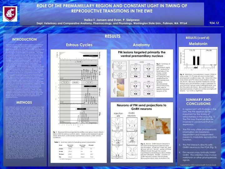
ROLE OF THE PREMAMILLARY REGION AND CONSTANT LIGHT IN TIMING OF REPRODUCTIVE TRANSITIONS IN THE EWE Heiko T. Jansen and Evan. P. Sleipness Dept. Veterinary and Comparative Anatomy, Pharmacology, and Physiology, Washington State Univ., Pullman, WA 99164 924.12 RESULTS RESULTS (cont’d) INTRODUCTION Melatonin Estrous Cycles Anatomy The seasonal cycle of reproduction in species such as sheep is thought to reflect the interaction among environmental signals, such as light, with brain regions responsible for generating reproductive cycles. Of particular importance for photoperiodic signaling is the pineal hormone melatonin, representing an endocrine signal for daylength . The highest concentration of melatonin binding sites is found in the pars tuberalis of the anterior pituitary (PT). The PT appears to play an important role in the photoperiodically-driven changes in prolactin secretion in sheep and in the generation of the endogenous circannual prolactin rhythm . Unlike the PT, concentrations of melatonin binding sites in the brain are much lower and relatively few specific brain regions mediating the physiological responses to melatonin have been identified. The premamillary region (PM) of the caudal hypothalamus in sheep contains a relatively high number of melatonin binding sites, when compared to surrounding brain regions. Furthermore, the PM responds directly to melatonin as revealed by the rise in LH concentrations in ewes with melatonin implants placed in the PM and maintained under long (inhibitory) daylengths (Malpaux et. Al., Endocrinology, 1998). Because the LH response observed in melatonin-implanted ewes is very similar to that seen following exposure to short daylengths, when melatonin secretion at night is prolonged, the results have been interpreted to indicate PM may serve as a neural site for photoperiodic integration. How this integration occurs with reproductive neuroendocrine centers is not known, nor is it known if the requirement for melatonin is absolute (i.e., is photoperiodic information conveyed in the absence of melatonin). Finally, we were interested in confirming whether the PM plays a role in the expression of photorefractoriness in the ewe as it does in the ram (Lincoln and Clarke, Soc. Study Fertil. Abst., 2000) and thereby implicate this brain region in the generation of circannual rhythms. PM lesions targeted primarily the ventral premamillary nucleus Fig. 2. Summary of PM lesion placements (right). Left - anatomical landmarks of the sheep caudal hypothalamus including PM and neighboring TM. Nissl staining and histidine decarboxylase immunoreactivity were used to identify histamine neurons of TM. Fig. 4. Melatonin concentrations (mean ± SEM) in ewes under 17L:7D (day 45) and constant light photoperiod conditions (day 135). Data from PMx ewes (bars) and PM-intact ewes (hollow circles) are shown. Because daytime melatonin concen-trations and concentrations under LL between Px and PM-intact groups were indistinguishable; only the PMx data are shown. Bars under each plot represent periods of lights-on (white) and lights-off (black). * p < 0.05 vs. nighttime SUMMARY AND CONCLUSIONS METHODS Neurons of PM send projections to GnRH neurons Adult, Suffolk-cross ewes (N=16) were housed outdoors at the Sheep Research Facility, University of Idaho, Latitude 47ºN, until March 1, 2001. Animals were fed alfalfa hay and provided mineral supplements with water available ad libitum. Five ewes remained outdoors and served as natural photoperiod controls. The remaining 11 ewes were moved indoors on March 1, 2001 into light-controlled photochambers. Photochambers were fitted with fluorescent fixtures to provide an illumination intensity of >300 lux at the level of the sheep’s head. All 11 ewes were initially exposed to a 90-day period of long-day (LD) photoperiod equivalent to that observed at the time of the summer solstice at this latitude (17h light:7h dark, 17L:7D) (Fig. 1). Lights were turned on at 0400h and turned off at 2100h with the aid of automatic digital timers. On May 30, 2001, the photoperiod for 7 of the 11 ewes was changed to constant light (24L:0D, LL) and these were maintained on LL for a period of 90 days. The remaining 4 ewes were maintained on LD for the duration of the experiment (330 days). After 180 days, the 7 ewes previously held under LL were then switched to a short-day (SD) photoperiod (9L:15D) for the duration of the experiment (150 days). from all ewes into heparinized tubes and centrifuged. Photochambers were provided with constant dim red illumination (<5lux) to facilitate nighttime blood sampling. On two separate occasions (day 45 and 135), jugular blood samples were collected from all animals housed in photochambers and at three different times of day: morning (0900h), mid evening (2300h), and late evening (0300h). Blood samples (5ml) were collected by jugular venipuncture twice weekly. The PM was defined initially based on histological material from similarly sized and perfused ewes. Based on this, the anatomical boundaries of the PM were defined as extending rostrally from the caudal boundary of the hypothalamic arcuate nucleus (Arc), 2.0mm lateral to the mamillary recess (mr) of the third ventricle and 1.5mm above the floor of the mamillary recess, and caudally to the mamillary bodies (Fig. 2). The PM was targeted bilaterally for destruction in 4 ewes (PMx) with the aid of a Radionics (Burlington, MA) radiofrequency lesion maker and electrode (tip diameter 2mm). The electrode was heated to a temperature of 60ºC for 60sec. In 3 additional ewes (PM-intact), the radiofrequency electrode was lowered to a brain region just dorsal to the target site; no temperature increase was induced. Serum progesterone and melatonin concentrations were determined by RIA. Pressure injections (150nl) of Biotinylated Dextran amine (10,000MW) were made into the PM and animals killed two weeks later. BDA was visualized using nickel-enhanced DAB as the chromagen and GnRH was visualized using the LR-5 antibody (generous gift of Dr. Robert Benoit; diluted 1:50,000)and unenhanced DAB. In agreement with findings in the ram, the PM appears to be important for the timing of refractoriness in the ewe (Fig. 1). The PM may therefore also be involved in circannual rhythm expression. The PM may utilize photoperiodic information via melatonin-dependent and independent means to modulate reproductive function. The PM interacts directly with GNRH neurons in the POA (Fig. 3). PM neurons may tonically inhibit GnRH. This inhibition may involve melatonin or other photoperiodic signals. GnRH neurons Injection site Fig. 1. Representative progesterone profiles and group means (black bars) ± SEM for estrous cyclicity data. Upper panel represents group mean data from control ewes maintained outdoors under natural conditions. Table 1. Summary data for breeding season parameters. Fig. 3. Above - GnRH neuron (brown) in the POA receiving inputs (arrows) from the PM region. Scale bar =10µm (A), 5µm (B) and 1µm (C). Far left - panels at three different levels through the caudal hypothalamus illustrating location of BDA injection (Red)site. Middle - Location of GnRH neurons (circles) that received BDA-positive inputs (red circles)from PM. Supported by USDA Animal Health Formula funds