Download
1 / 85
1.02k likes | 1.92k Vues
Intercalated BSc 2007-08. CELL DEATH an overview. Dr Cathy Baker 22 nd October 2007. How do cells die?. Killed by injurious agents Induced to commit suicide. NECROSIS. APOPTOSIS. LEARNING OBJECTIVES. Understand, describe and illustrate … Differences: necrosis vs. apoptosis

E N D
Intercalated BSc 2007-08 CELL DEATHan overview Dr Cathy Baker 22nd October 2007
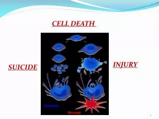
How do cells die? • Killed by injurious agents • Induced to commit suicide NECROSIS APOPTOSIS
LEARNING OBJECTIVES Understand, describe and illustrate … • Differences: necrosis vs. apoptosis • Morphological changes of apoptosis • Function of apoptosis • Principal biochemical mechanisms • Role of apoptosis in pathologies
Necrosis Apoptosis Lecture overview Function Morphological changes Biochemistry Pathology
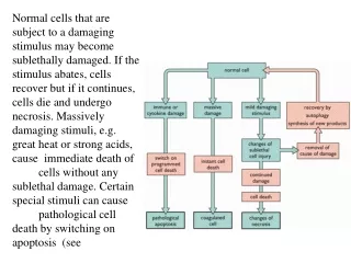
Necrosis • Mechanical injury & toxic agents • Cell groups • Membrane integrity destroyed • Cells and organelles swell, burst and leak contents • Inflammatory response • Other cells and tissues damaged
John Kerr et al Br.J.Cancer 26: 239-257, 1972
Apoptosis • Essential biological process • Cells have role in own death - told or decide to commit suicide • Programmed cell death (PCD)
Apoptosis • Distinct form of single cell death • Tightly regulated • Very localised • Energy consuming process • Membranes intact (early stages) • Safe disposal of cell corpse • No inflammation
Necrosis Apoptosis Morphological changes
Changes in cell morphology • Cells shrink and become detached from adjoining cells • Cytoskeleton collapses • Mitochondria remain intact • Plasma membrane develops bubbles (blebs) on surface
Nucleus and chromatin condense • Aggregates at periphery of nucleus • Nuclear envelope disintegrates • DNA fragmentation • Budding off and breakage into small membrane wrapped fragments - apoptotic bodies
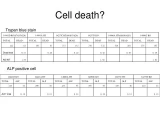
What happens to apoptotic cells and apoptotic bodies? • Ingested & degraded by phagocytes • Macrophages and dendritic cells • Adjacent cells in tissue • High speed and efficiency • Histologically inconspicuous • No inflammation
Necrosis Function Apoptosis Morphological changes
Function of apoptosis? • Deliberate removal of specific, unwanted cells • Organised and controlled manner • Without damaging other cells or tissues Circumstances?
Homeostasis • Constancy of internal environment • Tissue turnover • Cell numbers have to be maintained Homeodynamics
Embryonic development Removal of unwanted cells • Damage • Organ and tissue differentiation • Vestigial structures • Alteration of tissue form
5 weeks 8 weeks
Neurologicaldevelopment • Deletion of excess immature neurons that have failed to establish synaptic connections • Occurs in CNS and PNS • Prevents redundant cell in mature nervous system
Involution of tissue • Endometrial breakdown prior to menstruation • Regression of lactating breast tissue after weaning
Cell damage • Internal cell damage • Inappropriate 3o protein structure • Cell Infection • Viral • Stress • Starvation • DNA damage • Ionizing radiation, ROS
Necrosis Function Apoptosis Morphological changes Biochemistry
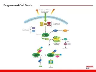
Biochemistry of apoptosis • Intense area of research • Complicated integrated mechanisms • Much more to be revealed! • Common core process • Underpins morphological changes • Four stage process
Stage 1- The Death Signal • Absence or withdrawal of positive survival factors • Presence of negative pro-apoptotic factors
Survival or positive signals • Cell survival relies receiving positive stimuli • Neuronal growth factor • Interleukin 2 for lymphocytes • Hormones • Withdrawal is a death signal • Default pathway for many cells
Death or negative signals • Signals to induce apoptosis • Damaged DNA • UV light and X rays • Chemotherapeutic drugs • Oxidants/free radicals • Oxidative stress • Death activators or receptor ligands
What are Death Activators? • Molecules that bind to specific receptors on cell surface • Tumour necrosis factor alpha • Lymphotoxin TNF beta • Fas ligand (CD95) • Binding of death activator to its specific receptor is a pro-apoptotic signal
Stage 2 - Integration and Transduction • Signals linked to execution phase through an integration stage • Uses positive and negative regulatory molecules • Inhibit, stimulate or forestall apoptosis
To die or not to die? Integrated balance between positive survival factors and negative death signals decides fate of cell
Common intracellular machinery for apoptosis The three main players • Family of enzymes - Caspases • Protein family - Bcl-2 proteins • Regulating gene - p53 gene
Caspases • Family of protease enzymes • 14 isoforms identified • Have Cysteine at active site • Synthesised as inactive precursors - procaspase • Not all involved in apoptosis
cleavage sites prodomain large subunits small subunits Procaspase structure
Initiator caspases Effector caspases Activated caspase has proteolytic activity
Activate other caspases • Amplify caspase activity Initiator caspases Apoptosis execution Effector caspases
Bcl-2 proteins • Large family of proteins • Named from B cell lymphoma • Some are pro-apoptotic some are anti-apoptotic
Bcl-2 proteins and apoptosis • Main mechanism is regulation of mitochondrial permeability • Cell survival stimuli induce the expression of anti-apoptotic Bcl proteins • Death signals induce pro-apoptotic Bcl proteins
p53 gene and p53 protein • p53 is tumour suppressor gene • Active gene product p53 produced in response to DNA and cell damage • Prevents cell completing cell cycle • If damage is minor - allows repair • If major - induces apoptosis • Complex mechanisms
Apoptotic transduction pathways • Mitochondrial or intrinsic pathway • Death activator or extrinsic pathway