Download
1 / 20
200 likes | 609 Vues
Anat 1 Chapter 8 : Articulations. Functional / Structural Classification of Joints. Synarthrosis (no movement) Bony Fusion (Synostosis) Fibrous (Suture and Gomphosis) Cartilaginous (Synchondrosis) Amphiarthrosis (little movement) Fibrous (Syndesmosis) Cartilaginous (Symphysis).

E N D
Functional / Structural Classification of Joints • Synarthrosis(no movement) • Bony Fusion (Synostosis) • Fibrous (Suture and Gomphosis) • Cartilaginous (Synchondrosis) • Amphiarthrosis(little movement) • Fibrous (Syndesmosis) • Cartilaginous (Symphysis) Give example location for each
3) Diarthrosis (free movement) • Always synovial joints • mono, di-, and triaxial • Strength vs. motility The greater the range of motion, the weaker the joint. • Dislocation = luxation • Partial dislocation = ? • “Double jointed”
Diarthroses = Synovial Joints • Have synovial cavity = space between two bones • Components that are always present (fig 8-1) • Components that are sometimes present
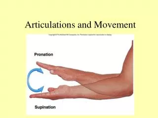
3 Types of Motion at Synovial Joints • Linear motion = gliding • Angular motion : • flexion, extension, hyperextension • ab-, adduction • circumduction • Rotation • left - right, internal or medial, external or lateral • supination, pronation
Special Movements Dorsiflexion, plantar flexion Protraction, retraction Elevation, depression Eversion inversion
6 types of Diarthroses • Gliding Joint • Hinge Joint • Pivot Joint • Ellipsoidal joint • Saddle joint • Ball & Socket joint
Gliding Joint articulating surfaces flat. • also found between carpals and tarsals • only slight movement - rotation prevented by ?
Hinge Joint Convex surface of bone 1 fits into concave surface of bone 2 • found in ? • monoaxial
Pivot Joint Projection of bone 1 articulates within ring of bone 2 Also found in proximal ends of ulna and radius pronation and supination rotation
Ellipsoidal (Condyloid) Joint Oval shaped condyle of bone 1 fits into elliptical cavity of bone 2 • Also found between phalanges & metacarpals/-tarsals • Angular motion in two planes (= )
Saddle Joint Articular surfaces shaped like saddle and rider Modified condyloid joint • Extensive angular motion without rotation • Also between malleus and incus
Ball and Socket Joint Ball like surface of bone 1 fits into cuplike depression of bone 2 • Found in ___________ • Allows for flexion, ab- or adduction and rotation ( _____axial)
Representative Articulations Temporomandibular Joint Mostly hinge joint, some gliding and rotation Articular disc
Intervertebral articulations Gliding joints between ____________________ Intervertebral discs: (Amphiarthroses) • annulus fibrosus:tough outer layer (fibrocartilage) • nucleus pulposus:soft, gelatinous core • Account for ~25% of vertebral column height – H2O loss during aging Intervertebral ligaments Fig 8-8
Disc Problems Fig 8-9 • Slipped disc vs. herniated disc • Most common sites for disc problems: • C5 - C6 • L4 - L5 • L5 - S1 • Lumbago • Laminectomy ( surgical removal vertebral arch by shaving laminae to access disc)
Glenohumeral Joint • Type? • Greatest range of motion (due to loose and shallow) • Most frequently dislocated • Stability provided by?
Hip Joint • Deep well fitted _______ joint • Participants ? • Stabilization: • Extracapsular and intracapsular ligaments (ligamentum teres = ligamentum capitis femoris) • Surrounding muscles • Most important normal movement? Fig 8-15
Knee Joint • Much more complex than elbow • Much less stable than other ______ • structurally 3 separate joints • Extra- and intracapsular ligaments • Locking of knee due to external rotation of tibia Figs 8-16 & 17
The end The end