Download
1 / 27
360 likes | 1.06k Vues
Coagulation / Coagulopathies. Hemostasis. Hemostasis is the ability of the body’s systems to maintain the integrity of the blood and blood vessels.

E N D
Hemostasis • Hemostasis is the ability of the body’s systems to maintain the integrity of the blood and blood vessels. • Hemostasis involves the arrest of the escape of blood by either natural (clot formation or vessel spasm), or by artificial (compression or ligation) means, or the interruption of blood flow to a part, or the artificial stimulation of clotting (i.e. electrocautery)
Hemostasis cont’d • Coagulation – the formation of a clot. • Platelets –aka thrombocytes. Small, anucleated, discoid-shaped, light blue staining cells that may have multiple, fine, ping to purple granules in the cytoplasm. Platelets are formed in the bone marrow. 1/3 of the total blood platelets are in the spleen, 2/3 remain in the circulating blood. Due to their adhesion and aggregation capabilities, platelets can occlude small breaks in blood vessels and prevent the escape of blood.
Hemostasis cont’d • Hemostasis involves a number of complex pathways, platelets, and coagulation (clotting) factors. • The ability for blood to clot, depends on vascular integrity, adequate numbers and normally functioning platelets, and a complete complement of coagulation factors. • ANY ALTERATION IN THESE PERAMETERS CAN RESULT IN A BLEEDING DISORDER!!
Coagulation Coagulation proceeds through a mechanical phase and a chemical phase. Each phase is counterbalanced by several thrombolytic factors to prevent excessive thrombosis.
Coagulation cont’d Mechanical Stage (Primary Hemostasis): • A blood vessel is ruptured or torn • The exposed vessel subendothelium is a charged surface, consisting of collagen fibers, and platelets are attracted to this surface. • Platelets congregate at the site of injury. • Platelets undergo morphologic and physiologic changes, causing the platelets to adhere to each other as well as the blood vessel endothelium. This causes an initial “plug” of platelets. • Platelets release the initiating factor for the chemical phase of hemostasis.
Coagulation cont’d Chemical Phase (Secondary Hemostasis)
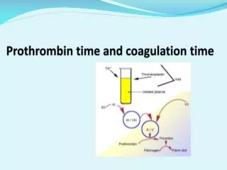
Coagulation cont’d Chemical Phase (Secondary Hemostasis) • Hemostasis relies upon a wide range of proteins called clotting factors and cofactors such as calcium and vitamin K. • Each factor participates in a chemical reaction that serves to initiate the next reaction in the pathway. • The end result of the coagulation cascade is the formation of prothrombin into thrombin which converts fibrinogen to fibrin. • Each clotting factor is essential for the formation of fibrin. • If any single factor is absent, the clotting cascade cannot be completed, and a fibrin mesh cannot be manufactured.
Coagulation cont’d Chemical Phase (Secondary Hemostasis) Fibrin gathers around the platelets , capturing a mesh of RBCs and WBCs to complete the clot http://www.youtube.com/watch?v=--bZUeb83uU&feature=related
The Formation of a Clot • Platets • Clotting Factors • Fibrinogen • Wbcs / Rbcs
Fibrinolysis • The final phase of hemostasis. • Involves the breakdown / degredation of the fibrin clot by the body.
So what happens if there is an interruption with the hemostatic process?
Coagulopathy : any disorder of blood coagulation.
Coagulation Testing • Evaluate specific portions of the hemostatic mechanisms. • Some tests measure the mechanical (primary) phase of hemostasis, while others can measure specific parts of the chemical phase. • All patients should be evaluated for coagulation defects before undergoing surgery.
Coagulation Testing cont’d • Blood samples should be collected carefully, with minimal tissue damage and minimal venous stasis. • Never collect samples through indwelling cathethers. • The preferred anticoagulant for coagulation test is sodium citrate.
Coagulation Test cont’d Platelet Count: manual platelet count is performed on a blood smear or on an automated analyzer. However, if a clotting disorder is suspected, a manual count is needed because the automated reader will misread clumped platelets. Platelets are counted at 400X in a hemacytometer.
Hemostatic Defects • Bleeding disorders may be caused by congenital or acquired defects in coagulation proteins, platelets, or the vasculature. • Most bleeding disorders found in veterinary species are secondary to another disease process.
Signs of hemostatic deficiencies in coagulation proteins usually involve delayed deep tissue hemorrhage and hematoma formation. • Signs associated with deficiencies include: Petechia Ecchymotic Hemorrhage
The majority if congenital coagulation factor disorders involve a deficiency or abnormality of a single factor. • Usually, signs of a congenital coagulopathy will be apparent before 6 mo of age.
Von Willebrand’s Disease • The most common inherited disorder of hemostasis. • Normally, von Willebrand’s factor (vWF) promotes platelet clumping. • With decreased amounts, or lack of this factor, a bleeding disorder results. • Affected breeds : Dobermans, GSD, Labs. • 54 breeds affected total in the U.S.
Von Willebrand’s Disease • Dogs with this disorder SHOULD NOT BE USED FOR BREEDING! • Predisposed breeds should be tested before breeding to ensure offspring will be clear. • Special care must be take with surgical procedures. • Clinical signs : • Easy bruising in predisposed breeds • Prolonged bleeding during estrus • Prolonged bleeding from venipuncture site
Secondary Coagulation Disorders • Result from decreased production or increased destruction of platelets. • Nutritional deficiencies • Liver disease • Ingestion of certain medications or toxic substances.
Secondary Coagulation Disorders cont’d • Thrombocytopenia refers to a decreased number of platelets and is the most common coagulation disorder seen in small animal veterinary practice. • The causes are often unknown. • Infection with certain bacterial, viral, and parasitic agents can result in thrombocytopenia
. • Thrombocytopenia can also occur as a result of bone marrow depression, which reduces the production of platelets or autoimmune disease that increases the rate of platelet destruction. • The liver is the site of production of most coagulation factors, any condition that affects liver function can result in a coagulation disorder.
Warafin Ingestion (Rat Bait) • Inhibits the funciton of vitamin K function. • Vitamin K is required for synthesis and activation of some coagulation factors. This creates a deficiency in several necessary components of the coagulation cascade.