Download
1 / 26
310 likes | 664 Vues
Assessment and Diagnosis of Pain Disorders. Pain Assessment: Goals. Characterize the pain Identify pain syndrome Infer pathophysiology Evaluate physical and psychosocial comorbidities Assess degree and nature of disability Develop a therapeutic strategy.

E N D
Pain Assessment: Goals • Characterize the pain • Identify pain syndrome • Infer pathophysiology • Evaluate physical and psychosocial comorbidities • Assess degree and nature of disability • Develop a therapeutic strategy
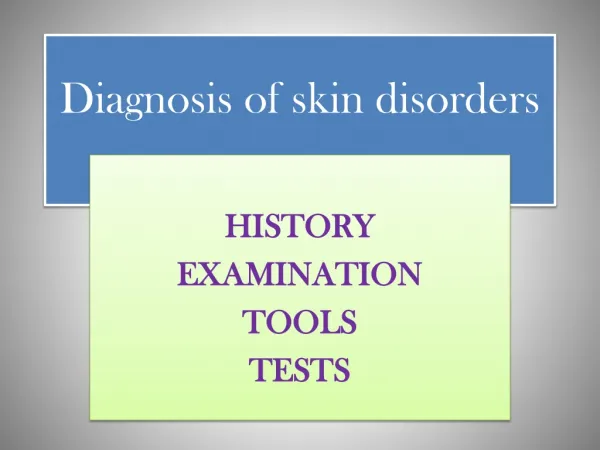
Comprehensive Pain Assessment • History • Physical examination • Appropriate laboratory and radiologic tests
Pain and Disability Nociception Other physical symptoms Physical impairment Neuropathic Psychologic Social isolation mechanisms processes Family distress Sense of loss or inadequacy Adapted with permission from Portenoy RK. Lancet. 1992;339:1026. Disability Pain
Pain History • Temporal features—onset, duration, course, pattern • Intensity—average, least, worst, and current pain • Location—focal, multifocal, generalized, referred, superficial, deep • Quality—aching, throbbing, stabbing, burning • Exacerbating/alleviating factors—position, activity, weight bearing, cutaneous stimulation
Nociceptive pain Neuropathic pain Idiopathic pain Psychogenic pain Commensurate with identifiable tissue damage May be abnormal, unfamiliar pain, probably caused by dysfunction in PNS or CNS Pain, not attributable to identifiable organic or psychologic processes Sustained by psychologic factors Pathophysiology
Pain Assessment Tools • Pain intensity scales • Verbal rating • Numeric scale • Visual analogue scale • Scales for children • Multidimensional pain measures • Brief Pain Inventory • McGill Pain Questionnaire
Acute pain Chronic pain Breakthrough pain Recent onset, transient, identifiable cause Persistent or recurrent pain, beyond usual course of acute illness or injury Transient pain, severe or excruciating, over baseline of moderate pain Pain Syndromes
Identify Pain Syndromes • Syndrome identification can direct assessment and predict treatment efficacy • Cancer pain syndromes • Bone pain • Pathologic fracture • Cord compression • Bowel obstruction • Noncancer-related pain syndromes • Atypical facial pain • Failed low-back syndrome • Chronic tension headache • Chronic pelvic pain of unknown etiology
Neuropathic Pain: Clinical Assessment • A comprehensive diagnostic approach to patients affected by neuropathic pain • Medical history • Examinations: general, neurologic, regional • Diagnostic workup: imaging studies, laboratory tests, nerve/skin biopsies, electromyography/nerve-conduction velocity (EMG-NCV) studies, selected nerve blocks
Medical History Ask patient about complaints suggestive of • Neurologic deficits: persistent numbness in a body area or limb-weakness, for example, tripping episodes, inability to open jars • Neurologic sensory dysfunction: touch-evoked pain, intermittent abnormal sensations, spontaneous burning and shooting pains
Neurologic and Regional Examinations In patients with neuropathic pain, examination should focus on the anatomic pattern and localization of the abnormal sensory symptoms and neurologic deficits
Diagnostic Workup: Lab Tests • Complete blood cell count with differential, erythrocyte sedimentation rate, chemistry profile • Thyroid-function tests, vitamin B12 and folate, fasting blood sugar, and glycosylated hemoglobin • Serum protein electrophoresis with immunofixation • Lyme titers, hepatitis B and C, HIV screening • Antinuclear antibodies, rheumatoid factor, Sjögren’s titers (SS-A, SS-B), antineutrophil cytoplasmic antibody
Diagnostic Workup: Lab Tests • Cryoglobulins • Antisulfatide antibody titers, anti-HU titers • Heavy metals serum and urine screens • Cerebrospinal fluid study for demyelinating diseases and meningeal carcinomatosis
Diagnostic Workup: Electrophysiologic Studies EMG-NCV and QST • To localize pain-generator/nerve or root lesion • To rule out • Axonal vs focal segmental demyelination • Underlying small-fiber or mixed polyneuropathy
Biopsies • Nerve (eg, sural nerve): to diagnose vasculitis, amyloidosis, sarcoidosis, etc. • Skin: to evaluate density of unmyelinated fibers within dermis and epidermis
Back Pain and Sciatica: Comprehensive Assessment • History • Medical • Psychosocial • Family • Previous trials • General examination • Musculoskeletal • Neurologic
Back Pain and Sciatica:Pain Assessment • Description • Duration • Intensity • Alleviating factors • Aggravating factors
Assessment of Patients With Low Back, Hip, and Leg Pain • Neurologic exam • DTRs, strength, sensitivity, gait • Regional exam of spine and leg • Inspection for scoliosis or skin rash, palpation for bone tenderness • Sciatic- and femoral-nerve stretching tests • SLR, reverse and contralateral SLR maneuver
Assessment of Patients With Low Back, Hip, and Leg Pain • Provocative mechanical joint tests • Truncal flexion for discogenic pain or spine instability • Truncal extension for facet joint disease • Patrick’s maneuver for hip disease (FABER test of both hips for SI joint disease)
Back Pain and Sciatica: Imaging Evaluation • Lumbosacral x-ray studies with flexion/ extension/oblique views • MRI of the spine • CT with 3-D reconstruction • CT plus myelography
Assessment of Chronic Back Pain and Sciatica: Diagnostic Blocks • Facet blocks to rule out facet joint pain • Provocative diskograms or disk blockade to rule out discogenic pain and pain associated with segmental spinal instability • Selective root blocks to determine location of root pain generator
Neoplasm Infection (diskitis, epidural abscess) Cauda-equina syndrome Compression Fx Assessment of Acute Back Painand Sciatica: “Red Flags” History Possible Diagnosis • Nighttime pain, fever, weight loss, history of cancer • Fever, IV drug abuse • Bladder, bowel dysfunction; leg weakness • Trauma
Back Pain and Sciatica MRI of the spine if patient demonstrates • “Red flags” • Neurologic deficits or progressive neurologic signs and symptoms • Pain persisting more than 6 wk
Headache Evaluation • History (duration, onset, frequency) • Is there a family history of headache? • Are there any known causes of headache? • What is the typical location(s)? • What does the pain feel like? • What makes it worse? • What makes it better? • What are the results of past evaluations? • Are there associated symptoms? Exam findings? • What is the patient’s sex?
Headache: Diagnostic Red Flags • Rash, meningeal signs, or fever • Onset after age 50 • Onset in a person with HIV or cancer • Abrupt onset • Worsening pain • Signs of focal neurologic disease