Download
1 / 13
130 likes | 620 Vues
In Vivo Noninvasive Serial Monitoring of FDG-PET Progression and Regression in a Rabbit Model of Atherosclerosis. Stephen G. Worthley, Zhuang Y. Zhang, Josef Machac, Gérard Helft, Cheuk Tang, Gary Y. H. Liew,

E N D
In Vivo Noninvasive Serial Monitoring of FDG-PET Progression and Regression in a Rabbit Model of Atherosclerosis Stephen G. Worthley, Zhuang Y. Zhang, Josef Machac, Gérard Helft, Cheuk Tang, Gary Y. H. Liew, Azfar G. Zaman, Matthew I. Worthley, Zahi A. Fayad, Monte S. Buchsbaum, Valentin Fuster, Juan J. Badimon Int J Cardiovasc Imaging, 1 Nov 2008, DOI 10.1007/s10554-008-9377-2 Copyright © 2008 Society for Heart Attack Prevention and Eradication. All Rights Reserved.
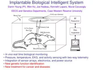
Introduction • Macrophages have a central role in atherosclerotic plaque inflammation and the development of an unstable plaque leading to fissuring and rupture. • Imaging techniques that can characterize plaque components or detect plaque inflammation may potentially be useful tools for risk stratification or monitoring of atherosclerotic disease. • We and others have shown the feasibility of positron emission tomography (PET) using [18F] fluorodeoxyglucose to assess macrophage content in atherosclerotic plaques. • Therefore, we sought to assess the progression and/or regression of macrophage content in atherosclerotic plaques in an experimental rabbit model using this noninvasive technique Copyright © 2008 Society for Heart Attack Prevention and Eradication. All Rights Reserved.
Methods: Experimental Model Atherogenic diet (n=8) Normal diet (n=4) 6 months 9 Months Atherogenic diet (n=4) W1 BD W13 BD PET 1 PET 2 • All 8 New Zealand White rabbits underwent induction: 9 months of atherogenic diet (0.2% Cholesterol) • All had balloon denudation of descending aorta at Week 1 and Week 13, utilizing a 4Fr Fogarty embolectomy catheter • Randomized to 6 months of normal diet (regression group) or continue same atherogenic diet (progression group) • FDG PET imaging at start and end of randomized period (Months 9 & 15) • All animals sacrificed after 2nd PET scan and histopathology of aorta performed Copyright © 2008 Society for Heart Attack Prevention and Eradication. All Rights Reserved.
Methods – FDG PET imaging • All procedures under general anesthesia after overnight fast • Prior to FDG PET, all rabbits underwent MRI for co-localization of anatomical structures (2D time of flight sequence). • For both PET & MRI, they were immobilized in a body-fitting thermosetting plastic holder to ensure exact positioning. • GE 2048-plus brain dedicated PET scanner; FDG dose: 1-2 mCi • PET imaging performed 30 mins after injection • Image co-registration with computer software of PET and MRI • FDG uptake expressed as ratio of aortic uptake-to-blood radioactivity as previously validated. • 25 consecutive axial slices (6 mm apart) from distal aortic arch were analyzed per animal. Pre & Post PET scans were matched in each. • Statistics: paired students t-test for matched aortic images pre & post dietary modification. Copyright © 2008 Society for Heart Attack Prevention and Eradication. All Rights Reserved.
Co-registration of PET & MRI • Demonstration of the co-registration of PET and MR images allowing the exact determination of FDG uptake in the aorta. • These images are from the same animal showing MRI (Panel A), FDG PET (Panel C) and the resultant fusion (Panel B). • The yellow arrows highlight the position of the aorta and the white arrows the position of the heart Copyright © 2008 Society for Heart Attack Prevention and Eradication. All Rights Reserved.
Results Serum total cholesterol • Baseline: 34±23 mg/dl End of induction: 949±141 mg/dl At end of randomized period: • Progression group: 936±340mg/dlRegression group: 27±6 mg/dl • Serum cholesterol increased significantly at end of induction and remained high in progression group but decreased in group fed with normal diet. Histopathology • Induction of 9 months atherogenic diet & balloon denudation resulted in significant thickening of arterial wall (lipid & fibrous). • Induced lesions were rich in macrophage areas as indicated by RAM-11 staining. Copyright © 2008 Society for Heart Attack Prevention and Eradication. All Rights Reserved.
Results - Histopatholgy • In vivo axial PET image showing the uptake of FDG in the thoracic aorta (black arrow) in a control rabbit (A), in a rabbit with mild atherosclerosis (B) and in a rabbit with more advanced atherosclerosis showing a higher uptake of FDG (C). • Corresponding histopathology stained with RAM-11 of A, B, C showing no macrophages (D) somemacrophages (F) and abundant macrophages (F) respectively Copyright © 2008 Society for Heart Attack Prevention and Eradication. All Rights Reserved.
FDG uptake in Progression Group • Box & Whisker plots showing all 100 matched aortic slices for 4 animals pre & post randomization in progression group. • Significant increase in aortic uptake-to-blood radioactivity after further 6 months of cholesterol feeding: 0.57±0.02 to 0.68±0.02, p=0.001 Copyright © 2008 Society for Heart Attack Prevention and Eradication. All Rights Reserved.
FDG uptake in Regression Group • Box & Whisker plots showing all 100 matched aortic slices for 4 animals pre & post randomization in regression group. • Significant decrease in aortic uptake-to-blood radioactivity after further 6 months of normal feeding: 0.67±0.02 to 0.53±0.02, p=0.001 Copyright © 2008 Society for Heart Attack Prevention and Eradication. All Rights Reserved.
Discussion • In this study, uptake of FDG was assessed in the same rabbit at two time points and a serial comparison between the same atherosclerotic aortic segments made for each rabbit. • We were able to monitor and identify significant changes in FDG uptake between the progression and regression groups. Although this may reflect serial changes in macrophage activity alone, we and others have previously shown FDG uptake to correlate with macrophage content. Ogawa, et al. J Nucl Med 2004; Zhang, et al. BMC Nucl Med 2006. • Studies have found disrupted plaque fibrous caps usually are heavily degraded and infiltrated by macrophages foam cells. These macrophage are activated, indicating ongoing inflammation at the site of plaque disruption. Lendon, et al. Atherosclerosis 1991; van der Wal, et al. Circulation 1994. Copyright © 2008 Society for Heart Attack Prevention and Eradication. All Rights Reserved.
Discussion • Others have shown in human carotid artery that symptomatic plaques have higher FDG PET activity than asymptomatic ones. They have also shown by autoradiography the location of macrophages to be at the fibrous cap. Rudd, et al. Circulation 2002 • Similarly, in human coronaries there is a correlation between plaque macrophage density and symptom severity in acute coronary syndromes. MacNeill, et al. JACC 2004 • Recently, a small study has demonstrated attenuation of plaque inflammation in humans with simvastatin utilizing FDG PET imaging. Tahara, et al. JACC 2006 Copyright © 2008 Society for Heart Attack Prevention and Eradication. All Rights Reserved.
Limitations • Using MRI to co-register FDG uptake has been validated previously, but potentially a PET/CT hybrid system could be more accurate. • Our study employed a small number of animals. • A cross over design may have added further to the potential of FDG PET in serial monitoring of changes in this model. • We have previously demonstrated atherosclerotic progression and regression utilizing high resolution MRI with histopathological correlation in the same animal model. Helft, et al. Circulation 2002 • We were not able to quantify plaque burden in this study as the MRI images were purely for co-localization. Copyright © 2008 Society for Heart Attack Prevention and Eradication. All Rights Reserved.
Summary • [18F]FDG-PET scanning in this animal model of atherosclerosis has allowed us to serially monitor macrophage content. • Thus FDG PET could potentially be used to serially monitor changes in atherosclerotic plaque macrophage content in response to therapies such as lipid lowering over time. Copyright © 2008 Society for Heart Attack Prevention and Eradication. All Rights Reserved.