Download
1 / 29
790 likes | 3.45k Vues
Interventional Radiology Coding Basics. PRESENTED BY:. Rosemary Waligorski, RHIT, CCS, RCC CHW IVR/CVIR Coding Compliance Spec . NvHIMA State Meeting April 24, 2010. Objectives and Goals. Basic understanding of interventional radiology Gain knowledge of key terms and phrases

E N D
Interventional Radiology Coding Basics PRESENTED BY: Rosemary Waligorski, RHIT, CCS, RCC CHW IVR/CVIR Coding Compliance Spec NvHIMA State Meeting April 24, 2010
Objectives and Goals • Basic understanding of interventional radiology • Gain knowledge of key terms and phrases • Practice – participate in case examples and exercises
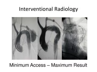
What is Interventional Radiology Interventional radiology is the non-surgical treatment using radiologic imaging, with contrast, to guide instruments (catheters, balloons, etc) through the body’s blood vessels and other organs. This type of procedure is done for both diagnostic and therapeutic procedures and is usually done on an outpatient basis. Some common types of procedures done in interventional radiology include: • Angiography • Angioplasty • Atherectomy • Stent insertion • Biopsies • Occlusion procedures
Interventional Radiology Terms • Angiography – Pictures taken of a blood vessel • Antegrade – with the flow • Bifurcation – a “splitting” or “forking” of vessels into two separate vessels • Bovine Arch – anomaly in the vessels coming off the aortic arch where the left common carotid actually comes off the right innominate artery instead of off the aorta • Catheter – instrument used in most percutaneous interventional radiology procedures • Contralateral – opposite side • First order – primary vascular branch
Interventional Radiology Terms (cont) • Great Vessels – those vessels that come off the aortic arch – in normal anatomy; the brachiocephalic, common carotid, and left subclavian • Guiding angiogram – imaging taken of vessel that the catheter is in so that the physician can verify where they’re at and where they still need to go • Ipsilateral – same side • Non-select catheter placement – catheter positioned in the aorta (from anywhere outside of the aorta) or placement in the access vessel itself without manipulation into another vessel
Interventional Radiology Terms (cont) • Retrograde – against the flow • Select catheter placement – catheter that is threaded: • Into a vessel off the vessel accessed • The aorta • Vena Cava • Vascular families – each separate set of vessels: • Off the vessel accessed • Off the Aorta • Off the Vena Cava
Some Simple Coding Rules • Code to the final catheter placement per vascular family • Do not code to where the wire is placed • Select catheter placement always takes precedence over non-select unless a second access is obtained • Code each vascular family separately • Code only one select catheter placement from each vascular family (36215-36217 & 36245-36247) • Make sure the proper codes are being used for catheter placement in regards to anatomy • Above the diaphragm 36215-36218 • Below the diaphragm 36245-36248
Key Words and Phrases to Look for • Roadmapping – this is NOT imaging • Verification of catheter placement is NOT imaging • In, engaged, cannulated, in the mouth of, in the orifice of, in the ostia; these describe the catheter being IN the vessel the physician is referring to or maybe getting ready to image • At, near, to, proximal to; these are words that describe the catheter NOT being in the vessel the physician is referring to
CPT Codes - Catheterization 36120 Introduction of needle or intracatheter; retrograde brachial artery 36140 extremity artery 36200 Introduction of catheter, aorta 36215 Selective catheter placement arterial system: each first order thoracic or brachiocephalic branch, within a vascular family 36216 initial second order thoracic or brachiocephalic branch, withina vascular family
CPT Codes – Catheterization (cont) 36217 initial third order or more selective thoracic or brachiocephalic branch, within a vascular family +36218 additional second order, third order and beyond, thoracic or brachiocephalic branch, within a vascular family (List in addition to code for initial second or third order vessel as appropriate)
CPT Codes – Catheterization (cont) 36245 Selective catheter placement, arterial system; each first order abdominal, pelvic, or lower extremity artery branch, within a vascular family 36246 initial second order abdominal, pelvic, or lower extremity artery branch, within a vascular family 36247 initial third order or more selective abdominal, pelvic, or lower extremity artery branch, within a vascular family +36248 additional second order, third order, and beyond, abdominal, pelvic, or lower extremity artery branch, within a vascular family (list in addition to code for initial second or third order vessel as appropriate)
CPT Codes - Radiology 75605 Aortography, thoracic, by serialography, radiological supervision & interpretation 75625 Aortography, abdominal, serialography, radiological supervision and interpretation 75630 Aortography, abdominal plus bilateral iliofemoral lower extremity, catheter, by serialography, radiological supervision and interpretation 75650 Angiography, cervicocerebral, catheter, including vessel origin, radiological supervision and interpretation 75660 Angiography, external carotid, unilateral, selective, radiological supervision and interpretation
CPT Codes – Radiology (cont) 75662 Angiography external carotid, bilateral, selective radiological supervision and interpretation 75665 Angiography, carotid cerebral, unilateral, radiological supervision and interpretation 75671 Angiography carotid cerebral, bilateral, radiological supervision and interpretation 75676 Angiography, carotid, cervical, unilateral, radiological supervision and interpretation 75680 Angiography, carotid, cervical bilateral, radiological supervision and interpretation
CPT Codes – Radiology (cont) 75685 Angiography, vertebral, cervical, and/or intracranial, radiological supervision and interpretation 75705 Angiography, spinal, selective, radiological supervision and interpretation 75710 Angiography, extremity, unilateral, radiological supervision and interpretation 75716 Angiography, extremity, bilateral, radiological supervision and interpretation
CPT Codes – Radiology (cont) 75722 Angiography, renal, unilateral, selective (including flush aortogram, radiological supervision and interpretation) 75724 Angiography, renal, bilateral, selective (including flush aortogram), radiological supervision and interpretation 75726 Angiography, visceral, selective or supraselective (with or without flush aortogram), radiological supervision and interpretation 75736 Angiography, pelvic, selective or supraselective, radiological supervision and interpretation +75774 Angiography, selective, each additional vessel studied after basic examination, radiological supervision and interpretation (list separately in addition to code for primary procedure
CPT Code 75774 • To code/charge CPT 75774, it is required that additional imaging beyond the basic exam is performed after an additional level of catheter selectivity is documented for the exam. • It is not appropriate to assign when images are obtained to “complete” the initial “runoff” exam, i.e. step-table, even if more selective catheter placement is performed. • Documentation of the clinical indication for needing to do so, e.g. poor visualization, occlusive disease, anatomical variants, etc., as well as imaging findings is also required.
Case Example Rules • Unless otherwise specified, all access is through the femoral artery • All findings are assumed reported unless otherwise stated
Catheter placement in aorta for abdominal aortogram Catheter placement in aorta for bilateral renal artery and abdominal aortogram Catheter placed in aorta and bilateral renal arteries for angiograms 4.Catheter placed in aorta for abdominal aortogram. Catheter then placed in the celiac trunk, inferior mesenteric artery, and superior mesenteric artery; contrast injected and images taken. Case Examples (Abdominal Aorta & Visceral)
Left femoral artery accessed, contrast injected for lower extremity angiography Left femoral artery accessed, catheter threaded up, over the abdominal aorta into the common iliac artery, contrast injected for angiography of the right lower extremity. Left femoral artery accessed; catheter threaded up into the aorta and placed at the bifurcation contrast injected for bilateral lower extremity angiography Femoral artery accessed. Catheter placed in contralateral common femoral artery for lower extremity angio. Catheter then pulled back to ipsilateral common iliac and contrast injected for lower extremity angio Case Examples (Lower Extremity)
Catheter threaded into the right subclavian for upper extremity angiography Catheter placed in the right brachiocephalic artery and the left subclavian artery. Upper extremity angiography performed Access in the right retrograde brachial artery. Upper extremity angio performed. Access in the right brachial artery for upper extremity angiography. Catheter then threaded up, into, and over the aortic arch and into the left subclavian for upper extremity angiography. Case Examples (Upper Extremity)
Catheter placed in the aortic arch and aortogram performed showing patent origins of the right brachiocephalic, left common, and left subclavian vessels. Slight tortuosity seen in the arch itself. Catheter placed in the aortic arch. Contrast is injected and angiography is taken of the bilateral common carotids and bilateral internal carotids. Same as number 14 with the addition of the external carotid arteries Case Examples (Head & Neck)
Catheter threaded through the aortic arch and placed in the right and left internal carotid arteries and the left vertebral artery for angiography Catheter placed in the right vertebral, right internal carotid, left internal carotid for angiography. Catheter placed in the left subclavian for angio of left vertebral. Catheter placed in the bilateral common carotid arteries for angiography. Then the catheter was placed in the bilateral external and internal carotid arteries for angiography. Catheter placed in: right vertebral, right external & internal carotids, left internal and external carotids, and left vertebral arteries where imaging of all these vessels were done and images reported. Case Examples (Head & Neck cont)
Case Examples (cont) 20. Local anesthesia provided to patient before procedure began. Right common femoral artery was prepped and draped in a sterile fashion then accessed with small nick incision. Multiple attempts were made to advance the wire and catheter but because of the occluded right external iliac artery, catheters were withdrawn and attention turned to the left groin. Using the same technique as the right groin, access into the left femoral artery was obtained. Catheter was then threaded up into the abdominal aorta to the level of the renal arteries. Aortography was performed with run-off down to the bilateral common femorals Images were obtained, after which all catheters were withdrawn and hemostasis obtained through manual pressure.
Case Examples (cont) 21. From R groin access, an abdominal aortogram is performed via cath placement in the abdominal aorta and injection above the renal arteries w/ findings documented followed by BLE angiography via cath movement to the bifurcation and injection revealing an abnormality of the R popliteal artery. The catheter is “pulled back” into the R femoral artery and additional injection or imaging is performed of the popliteal artery.
Case Examples (cont) 22. Utilizing Seldinger technique and 1% Xylocaine for local anesthesia, a #5 French pigtail catheter was inserted via the right common femoral artery and advanced into the aortic arch where arch aortography was performed. Bilateral carotid arteriograms and bilateral vertebral arteriograms were performed after selective catheterization. The following catheters were utilized: JB2 in the right common carotid, Simmons I in the left common carotid, and a #5 French vertebral catheter in both vertebral arteries. The catheters were removed and hemostasis obtained. The patient tolerated the procedure well. There were no apparent complications (cont)
Case Examples (cont) • Arch aortography: Shows normal orientation of the great vessels , with the left common carotid notably tortuous proximally. No other focal abnormalities are seen. • Right carotid arteriogram: There is approximately 40% narrowing of the junction of the right common carotid with the internal carotid from a small posterior located plaque. Otherwise, the right extracranial internal carotid appears unremarkable. The right external carotid appears unremarkable. There is noted a 7 x 6.5 mm aneurysm originating form the distal internal carotid near the level of the origin of the ophthalmic artery. The aneurysm projects medially and posteriorly. The right anterior and middle cerebral arteries appear unremarkable. • Left carotid arteriogram: No focal abnormalities are seen. No extracranial or intracranial abnormalities are evident. No aneurysms are identified. • Left vertebral arteriogram: No focal abnormalities are seen. No aneurysm is evident. The basilar tip is unremarkable. No aneurysms are seen around the region of the posterior inferior cerebellar arteries. • Right vertebral arteriogram: No focal abnormalities are identified. No aneurysms are seen. The basilar tip is unremarkable
36200, 75625 36200, 75625 36245, 36245, 75724 36215, 36215, 36215, 75726, 75726, 75726 36140, 75710 36245, 75710 36200, 75716 36246, 75716 36216, 75710 36215, 36215, 75716 36120, 75710 36215, 75710 36200, 75650 36200, 75680, 75671 36200, 75680, 75671 36217, 36216, 36216, 75671, 75685, 75685 36217, 36218, 36216, 36215, 75671, 75685, 75685 36217, 36218, 36216, 36218, 75680, 75671, 75662 36217, 36218, 36218, 36216, 36218, 36216, 75662, 75671, 75680, 75685, 75685 36140, 36200, 75630 36200, 75625, 75716 36217, 36218, 36215, 36216, 75650, 75671, 75680, 75685, 75685 Answers to Case Examples
Thank You rosemary.waligorski@chw.edu