Download
1 / 35
360 likes | 1.02k Vues
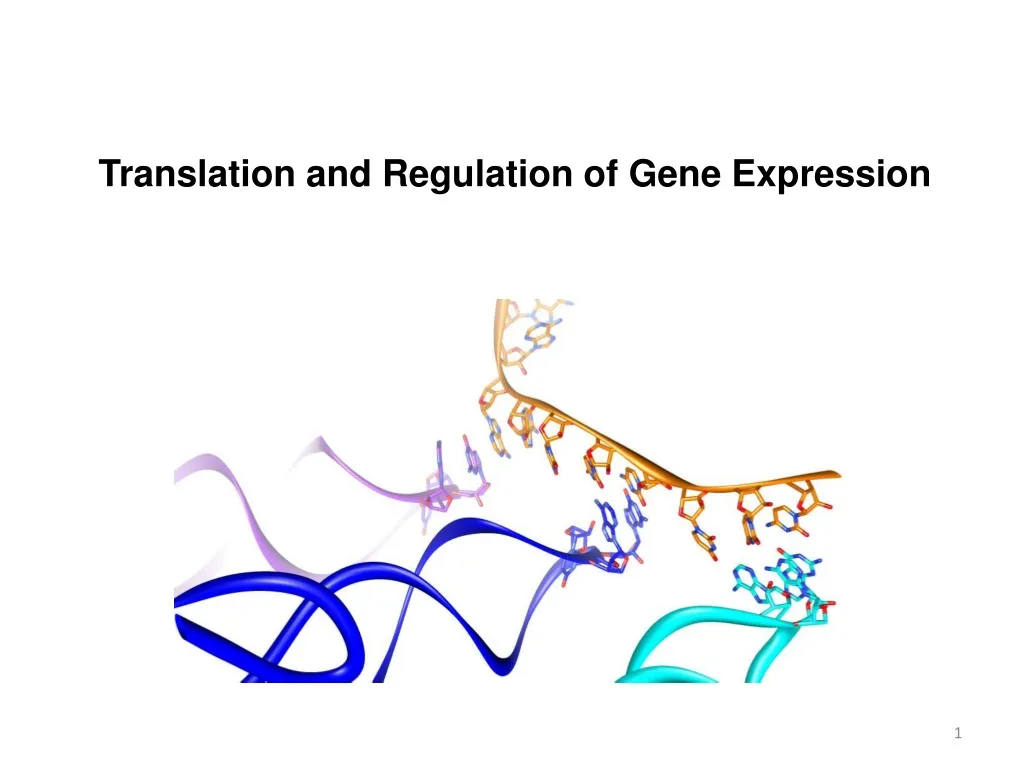
Translation and Regulation of Gene Expression. tRNA Tertiary Structure. Forms of DNA Helices. Continuous A-Form helix formed by Acceptor and T Y C-stems. L-shaped structures with ~60 Å legs. Helical junction is stabilized by several absolutely conserved base pairs.

E N D
tRNA Tertiary Structure Forms of DNA Helices Continuous A-Form helix formed by Acceptor and TYC-stems L-shaped structures with ~60 Å legs Helical junction is stabilized by several absolutely conserved base pairs Anticodons are ‘looped out’
Aminoacyl-tRNA Synthase Forms of DNA Helices Every amino acid has at least one aaRS Anticodon Loop Amino Acid Binding Site Matching the amino acid to a specific anticodon is the role of the aaRS. Must be sensitive to the flexibility of wobble position
Incoming Amino Acid Activation Forms of DNA Helices Amino acid is originally activated by ATP to form an aminoacyl-AMP intermediate Incoming tRNA displaces AMP to form an aminoacyl-tRNA tRNA
The Ribosome Forms of DNA Helices 2.5 MDa complex (in E. coli) Consists of 3 RNA chains 16S small subunit 23S + 5S large subunit and 52 proteins 21 associated with small subunit 31 associated with large subunit Sedimentation coefficient (Svedberg Units) Reports on the size and hydrodynamic properties of a particle
Aminoacyl-tRNA Binding to the Ribosome Forms of DNA Helices 30S subunit binds mRNA while 50S anchors tRNAs and catalyzes the peptide chain elongation • 3 distinct tRNAs binding sites • A site Aminoacyl site • binds incoming aa-tRNA • P site Peptidyl-transfer Site • tRNA with growing peptide chain • E site Exit Site • tRNA bound that lacks peptide and weakly base paired with mRNA
Aminoacyl-tRNA Binding to the Ribosome Forms of DNA Helices • A site Aminoacyl site • binds incoming aa-tRNA • P site Peptidyl Site • tRNA with growing peptide chain • E site Exit Site • tRNA bound that lacks peptide and weakly base paired with mRNA
Peptide Elongation at the Ribosome Forms of DNA Helices
Translation Initiation Forms of DNA Helices Orientation of mRNA on the ribosome The 16S rRNA contains a purine rich sequence that recognizes a pyrimidine rich sequence ~10 nucleotides downstream from the start codon – Shine Dalgarno Sequence Specialized tRNA for initiation The initial tRNA to insert into the P-site is a formaldehyde modified tRNA
Peptide Elongation at the Ribosome Forms of DNA Helices Chain Elongation happens in 3 steps Decoding – ribosomes selects and binds the proper tRNA Transpeptidation – peptide group on the P-site tRNA is transferred to the A-site tRNA Translocation – A-site tRNA transferred to the P-site and P-site tRNA transferred to the E-site
Translation Initiation Forms of DNA Helices • Translation Initiation is a complex process: • 30S and 50S subunits are initially separated • IF-3 keeps them from reassembling • mRNA binds to the 30S subunit (guided insertion) • fMet modified tRNA in a complex with IF-2 binds to 30S • This is assisted by IF-1
Translation Initiation Forms of DNA Helices
Translation Initiation in Eukaryotes Forms of DNA Helices eIF = eukaryotic initiation factors • eIF4E interaction with 5’cap • 2 Trp present planar groove for m7G to intercalate • H-bonds localized to W-C face and 5’ PP
Peptide Elongation at the Ribosome Forms of DNA Helices Decoding Process EF-Tu forms a complex with GTP (displacing EF-Ts) and the incoming aa-tRNA This complex binds to the empty mRNA at the A Site (energy dependent step) EF-Ts displaces the spent GDP Translocation Process The A-site tRNA displaces the P-site (uncharged) tRNA in an energy dependent process EF-G is the GTPase that hydrolyzes the GTP to promote translocation
Peptide Termination Forms of DNA Helices
Transcriptional Repression Forms of DNA Helices Promoter/Operator Not all genes need to be ‘turned on’ at all times. Doing so would be very wasteful Organisms have evolved mechanisms to control the cellular levels of proteins in the cell Inducer
Common Features of Regulatory Proteins Forms of DNA Helices • The recognition sequences tend to be palindromic • Regulatory proteins tend to interact as symmetric dimers (or dimers of dimers) • Base specific contacts are not always obvious • Intricate water networks • In bacteria, the helix-turn-helix motif (HTH) is very common • Variations: Winged Helix-Turn-Helix
Transcriptional Repression Forms of DNA Helices
Lac Repressor Forms of DNA Helices
Transcriptional Regulation – Deviant Promoters Forms of DNA Helices
Transcriptional Regulation – Deviant Promoters Forms of DNA Helices Suboptimal spacing or sequence of the Sigma Factor recognition elements decreases transcription efficiency CATATCGCTTGACTCCGTACATGAGTACGGAAGTAAGGTTACGCTAT -35 -10
Transcription Activators – MerR Family Forms of DNA Helices Sigma factor recognition elements are on opposite faces of the DNA helix CATATCGCTTGACTCCGTACATGAGTACGGAAGTAAGGTTACGCTAT To increase transcription efficiency: the spacing of the -10 and -35 elements must be changed OR We need another sigma factor OR We need to add another recognition element that increases affinity of RNAP for promoter Why won’t this work?
Catabolite Activator Protein (CAP) Forms of DNA Helices cAMP levels are inversely proportional to glucose levels cAMP When glucose, an organisms primary energy source, is low, a large array of catabolic enzymes are produced CAP controls the expression of over 100 genes that play a role in sugar metabolism CAP functions primarily to aid poor promoters HOW?
Catabolite Activator Protein (CAP) Forms of DNA Helices cAMP bound in activator domain a subunit makes contact with DNA and CAP cAMP-CAP promotes binding to the a subunit of RNAP CAP is a symmetric dimer that senses cAMP concentrations in in the Activation/Dimerization domain and binds to palindromic DNA by a wingedHTH motif in the DNA Binding Domain
Catabolite Activator Protein (CAP) Forms of DNA Helices
Transcription Activators - MerR Forms of DNA Helices MerR family proteins are symmetric dimers of HTH motifs connected by a single dimerization helix DNA binding occurs through a double winged HTH motif Primary interaction occurs through the HTH in the major groove Secondary interactions (primarily backbone) occur through the b-haripin that forms a wing as well as a second wing formed by 2 sequential helices
Transcription Activators - MerR Forms of DNA Helices Helix bending by DNA binding proteins is not particularly uncommon This family is unique among these proteins due to the twisting of the helix in addition to bending. Twisting is accomplished by ripping apart two base pairs make the minor groove significantly wider, and more solvent exposed
Post-transcriptional Regulation: microRNA Forms of DNA Helices
Post-transcriptional Regulation: Riboswitches Forms of DNA Helices
Regulation of the trp Operon Forms of DNA Helices
Regulation of the trp Operon – Attenuator Structure Forms of DNA Helices Leader Sequence = 5’ end of mRNA that is transcribed Two possible conformations of the attenuator sequence Spontaneous termination of transcription A terminator stem loop forms that causes the RNA polymerase to stall and “fall off” the mRNA
Regulation of the trp Operon – Attenuator Structure Forms of DNA Helices Leader Sequence = 5’ end of mRNA that is transcribed Two possible conformations of the attenuator sequence TrpTrp dimer (unstructured) More stable conformation (lots of GC stacking) Alternate structure (“antiterminator”) requires some way to prevent the formation of the more stable stem loops Trp makes up < 1% of amino acids in proteins – the presence of this ditryptophan is very unlikely A terminator stem loop forms that causes the RNA polymerase to stall and “fall off” the mRNA
Regulation of the trp Operon Forms of DNA Helices Ribosome plays an important role! Video Summary: https://www.youtube.com/watch?v=YAr18UR2dos • High [Trp] • Plenty of tRNA-Trp • Ribosome sequesters regions 1 and 2 • Termination Stem loop stable • Low [Trp] • Limited tRNA-Trp • Ribosome stalls at region 1 • Region 2 available for 2-3 stem loop formation (“antiterminator”) • No Terminator Not enough tRNA-Trp
Overview Forms of DNA Helices https://www.youtube.com/watch?v=u9dhO0iCLww