Download
1 / 10
100 likes | 119 Vues
The aim of the present study was undertaken to evaluate the effects of a Consciousness Energy Healing (The Trivedi Effect®) Treatment based test formulation on skin health. The test formulation and DMEM were separated into two parts. One part of each received the Consciousness Energy Healing Treatment by Shirley Theresa Holmlund and were termed as the Biofield Energy Treated samples, while the other parts were considered as the untreated test samples. MTT assay showed more than 70% cells were viable in all the tested concentrations in three cell lines and that the test formulation was safe and nontoxic. The percent cell proliferation using BrdU assay was significantly increased by 343.57%, 100.29%, and 132.6% in the UT-DMEM BT-Test formulation, BT-DMEM UT-Test formulation, and BT-DMEM BT-Test formulation groups, respectively at 8.75 µg/mL compared to the UT-DMEM UT-Test formulation group. The collagen synthesis was significantly increased by 5.94% and 10.84% in the UT-DMEM BT-Test formulation and BT-DMEM BT-Test formulation groups, respectively at 0.625 µg/mL with respect to the untreated group. Elastin level was significantly (p≤0.001) increased by 15.95%, 58.31%, and 47.18% at 2.5 µg/mL in the UT-DMEM BT-Test formulation, BT-DMEM UT-Test formulation, and BT-DMEM BT-Test formulation groups, respectively compared to the untreated group. The level of hyaluronic acid was significantly increased by 15.13% and 68.56% in the BT-DMEM UT-Test formulation and BT-DMEM BT-Test formulation groups, respectively at 0.625 µg/mL compared to the untreated group. The level of melanin was significantly reduced by 17.06%, 6.94%, and 13.07% in the UT-DMEM BT-Test formulation, BT-DMEM UT-Test formulation, and BT-DMEM BT-Test formulation groups, respectively at 0.063 µg/mL compared to untreated group. The effect of skin protection upon exposure with UV-B rays displayed some alteration of the percent cell viability in all the tested groups. The wound healing activity by scratch assay exhibited significant wound closure and cell migration in the test formulation and DMEM in HFF-1 cells compared to the untreated group. Altogether, the data suggests that the Consciousness Energy Healing Treated DMEM and test formulation exhibited better skin protective responses compared to the untreated DMEM and test formulation group with respect to the skin health parameters. Therefore, the Biofield Energy Treated test formulation could be developed as a potential cosmetic product against various skin problems viz. eczema, vitiligo, psoriasis, acne, hives, rosacea, ichthyosis, seborrheic dermatitis, erythema, contact dermatitis, skin aging, wrinkles, etc. | Authors:<br>Mahendra Trivedi, Dahryn Trivedi, Alice Branton, Gopal Nayak

E N D
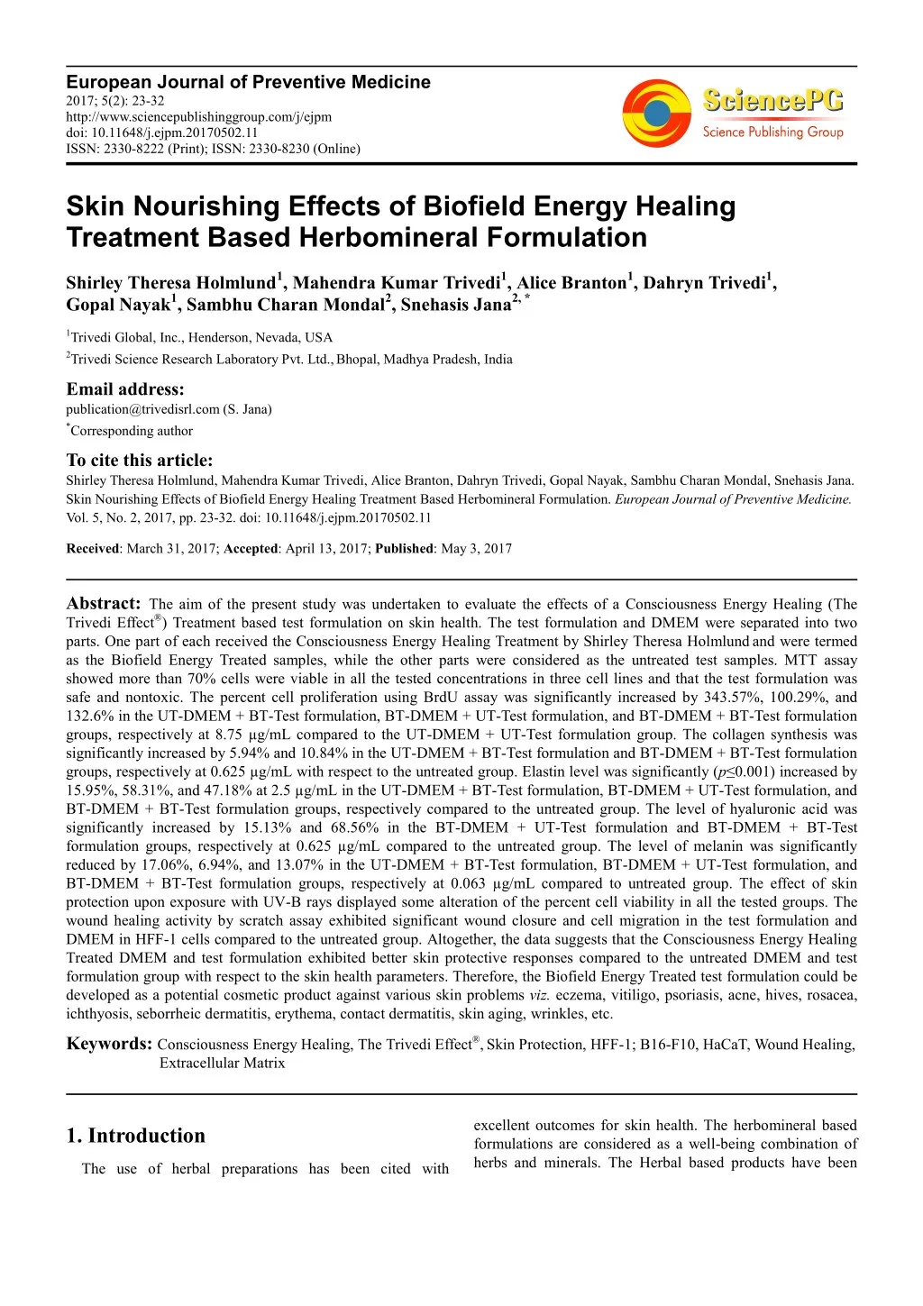
European Journal of Preventive Medicine 2017; 5(2): 23-32 http://www.sciencepublishinggroup.com/j/ejpm doi: 10.11648/j.ejpm.20170502.11 ISSN: 2330-8222 (Print); ISSN: 2330-8230 (Online) Skin Nourishing Effects of Biofield Energy Healing Treatment Based Herbomineral Formulation Shirley Theresa Holmlund1, Mahendra Kumar Trivedi1, Alice Branton1, Dahryn Trivedi1, Gopal Nayak1, Sambhu Charan Mondal2, Snehasis Jana2, * 1Trivedi Global, Inc., Henderson, Nevada, USA 2Trivedi Science Research Laboratory Pvt. Ltd.,Bhopal, Madhya Pradesh, India Email address: publication@trivedisrl.com (S. Jana) *Corresponding author To cite this article: Shirley Theresa Holmlund, Mahendra Kumar Trivedi, Alice Branton, Dahryn Trivedi, Gopal Nayak, Sambhu Charan Mondal, Snehasis Jana. Skin Nourishing Effects of Biofield Energy Healing Treatment Based Herbomineral Formulation. European Journal of Preventive Medicine. Vol. 5, No. 2, 2017, pp. 23-32. doi: 10.11648/j.ejpm.20170502.11 Received: March 31, 2017; Accepted: April 13, 2017; Published: May 3, 2017 Abstract: The aim of the present study was undertaken to evaluate the effects of a Consciousness Energy Healing (The Trivedi Effect®) Treatment based test formulation on skin health. The test formulation and DMEM were separated into two parts. One part of each received the Consciousness Energy Healing Treatment by Shirley Theresa Holmlundand were termed as the Biofield Energy Treated samples, while the other parts were considered as the untreated test samples. MTT assay showed more than 70% cells were viable in all the tested concentrations in three cell lines and that the test formulation was safe and nontoxic. The percent cell proliferation using BrdU assay was significantly increased by 343.57%, 100.29%, and 132.6% in the UT-DMEM + BT-Test formulation, BT-DMEM + UT-Test formulation, and BT-DMEM + BT-Test formulation groups, respectively at 8.75 µg/mL compared to the UT-DMEM + UT-Test formulation group. The collagen synthesis was significantly increased by 5.94% and 10.84% in the UT-DMEM + BT-Test formulation and BT-DMEM + BT-Test formulation groups, respectively at 0.625 µg/mL with respect to the untreated group. Elastin level was significantly (p≤0.001)increased by 15.95%, 58.31%, and 47.18% at 2.5 µg/mL in the UT-DMEM + BT-Test formulation, BT-DMEM + UT-Test formulation, and BT-DMEM + BT-Test formulation groups, respectively compared to the untreated group. The level of hyaluronic acid was significantly increased by 15.13% and 68.56% in the BT-DMEM + UT-Test formulation and BT-DMEM + BT-Test formulation groups, respectively at 0.625 µg/mL compared to the untreated group. The level of melanin was significantly reduced by 17.06%, 6.94%, and 13.07% in the UT-DMEM + BT-Test formulation, BT-DMEM + UT-Test formulation, and BT-DMEM + BT-Test formulation groups, respectively at 0.063 µg/mL compared to untreated group. The effect of skin protection upon exposure with UV-B rays displayed some alteration of the percent cell viability in all the tested groups. The wound healing activity by scratch assay exhibited significant wound closure and cell migration in the test formulation and DMEM in HFF-1 cells compared to the untreated group. Altogether, the data suggests that the Consciousness Energy Healing Treated DMEM and test formulation exhibited better skin protective responses compared to the untreated DMEM and test formulation group with respect to the skin health parameters. Therefore, the Biofield Energy Treated test formulation could be developed as a potential cosmetic product against various skin problems viz. eczema, vitiligo, psoriasis, acne, hives, rosacea, ichthyosis, seborrheic dermatitis, erythema, contact dermatitis, skin aging, wrinkles, etc. Keywords: Consciousness Energy Healing, The Trivedi Effect®,Skin Protection, HFF-1; B16-F10, HaCaT, Wound Healing, Extracellular Matrix excellent outcomes for skin health. The herbomineral based formulations are considered as a well-being combination of herbs and minerals. The Herbal based products have been 1. Introduction The use of herbal preparations has been cited with
24 Shirley Theresa Holmlundn et al.: Skin Nourishing Effects of Biofield Energy Healing Treatment Based Herbomineral Formulation used as holistic medicine for thousands of years, however the modern populations are dependent on various other forms like drugs, cosmetics, and agrochemicals [1]. These types of formulations also have been reported therapeutic benefits against different chronic diseases. Most of the cosmetic preparations used against skin darkening activity are based on the presence of phytoconstituents, which are derived from herbs [2], and their high demands are due to lower incidence of adverse effects compared with the synthetic compounds [3, 4]. Additionally, various types of products like photo- aging, antioxidant (i.e. vitamin B3, C, and E), invasive measures (gene therapy and chemical peels) and several devices (laser energy, injectable, etc.) are used for skin health and rejuvenation [5, 6]. Based on the retrieved scientific literature, the proprietary herbomineral formulation consists of essential minerals (zinc chloride, sodium selenate, and sodium molybdate), vitamin tetrahydrocurcumin (THC), and herbal extract (Centella asiatica). Each ingredient already has been proven for its potential activity on skin health as evidenced through use in various medicines as well as cosmeceuticals. Zinc is an essential cofactor of various metalloenzymes and it protects the skin from UV irradiation and has been used as a destructive agent for the management of cancer [7, 8]. Sodium selenate enhances the repair system of DNA segments and reduces the risk of new cancer development [9, 10]. Numerous literature suggests that it prevents skin cancers in combination with L-selenomethionine [11, 12]. Molybdenum is also an essential element for humans, animals, and plants and acts as a key constituent of various important enzymes [13, 14]. Ascorbic acid plays a vital role in repair of damaged skin and modulates collagen production [15]. THC exhibits antioxidant property and has been routinely used as a skin care formulation for the treatment of various skin related problems [16, 17]. The herbal extract of C. asiatica can enhance the process of wound healing and provides significant benefits in skin care products. Hashim et al. (2011) reported that C. asiatica leaves extract enhanced the synthesis of collagen and has the potential antioxidant, anti-cellulite, and UV protectant activities. It is also used in proprietary medicinal products for the treatment of cutaneous ulcer, hypertrophic scars, keloids, and wound healing disorders [18-20]. The National Center for Complementary and Integrative Health (NCCIH), allows the use of Complementary and Alternative Medicine (CAM) therapies like Biofield Energy as an alternative treatment in the healthcare field. About 36% of US citizens are regularly using some form of CAM [21], in their daily life. CAM embraces numerous Energy Healing Therapies; Biofield Therapy is one of the energy medicines used worldwide to improve overall human health. Researchers have shown that short-lived electrical events or action potential exists in the several types of mammalian cells such as neurons, muscles, and endocrine cells [22]. For instance, when the cells present in central nervous system of human body communicate with each another by means of electrical signals that propagate along the nerve impulses. Therefore, it was hypothesized that Biofield exists around the human body and evidence electromyography, electrocardiography electroencephalogram [23]. Thus, a Biofield Energy Healing Practitioner has the ability to harness the energy from the environment and can transmit it into any object (living organism or non-living material) around the globe. The object(s) always receives the energy and respond it in a useful way that is called “Biofield Energy”. This process is known as “Biofield Energy Healing”. Biofield Energy Healing has been approved as an alternative method that has an impact on various properties of living organisms in a cost- effective manner [24, 25]. The Trivedi Effect® Unique Biofield Energy Healing Treatment has been known to alter the response in a wide-spectrum fields in living and non- living systems viz. materials science [26-28], agriculture [29, 30], microbiology [31-33] biotechnology [34, 35]. Based on the excellent outcome of the Biofield Energy Treatment, the authors designed this study to investigate the impact of the Biofield Energy Healing Treatment on DMEM and the test formulation with respect to various skin health parameters using three cell lines such as human foreskin fibroblast (HFF-1), human keratinocytes (HaCaT), and mouse melanoma (B16-F10) cell lines. was found using and (L-ascorbic acid), 2. Materials and Methods 2.1. Chemicals and Reagents Fetal bovine serum (FBS) and Dulbecco's Modified Eagle's Medium (DMEM) were purchased from Gibco, USA. Antibiotics solution (penicillin-streptomycin) was procured from Himedia, India, while 3-(4, 5-diamethyl-2-thiazolyl)-2, 5-diphenyl-2H-tetrazolium) (MTT), Direct Red 80 and ethylene diamine tetra acetic acid (EDTA) were purchased from Sigma, USA. L-ascorbic acid was purchased from Alfa- Aesar, while kojic acid was purchased from Sigma, USA. Epidermal growth factor (EGF) was procured from Gibco, ThermoFisher, USA. ELISA kits were procured from CUSABIO and CusAb Co. Pvt. Ltd., USA. Zinc chloride was purchased from TCI, Japan, sodium selenate from Alfa- Aesar, USA, while sodium molybdate from Sigma-Aldrich, USA. Tetrahydrocurcumin and Centella asiatica extract were procured from Novel Nutrients Pvt. Ltd., India and Sanat Products Ltd., India, respectively. All the other chemicals used in this experiment were analytical grade procured from India. 2.2. Cell Culture The human foreskin fibroblast-1 (HFF-1) cells were procured from American Type Culture Collection (ATCC), USA. B16-F10 (mouse melanoma) cells were procured from National Centre for Cell Science (NCCS), Pune. HFF-1 and B16-F10 cell lines were maintained in the growth medium DMEM supplemented with 15% FBS, with added antibiotics penicillin (100 U/mL) and streptomycin (100 µg/mL). The growth condition of cell lines were 37°C, 5% CO2, and 95%
European Journal of Preventive Medicine 2017; 5(2): 23-32 25 humidity. L-ascorbic acid (for ECM, UV-B protection, and wound healing assay) in concentrations ranges from 10 µM to 1000 µM, while kojic acid (for melanin synthesis) concentrations ranges from 1 mM to 10 mM, FBS (0.5%) was used in cell proliferation (BrdU) assay, while EGF 10 µM was used in MTT assay. each well was read at 540 nm using Synergy HT micro plate reader, BioTek, USA. The concentrations exhibiting % cytotoxicity of < 30 % was considered as non-cytotoxic [36, 37]. The percentage of cell viability was calculated using formula (1): % Cell viability = (X*100)/R) (1) 2.3. Experimental Design Where, X represents the absorbance of the cells corresponding to positive control and test groups and R represents the absorbance of the cells corresponding to the baseline (control cells) group. The experimental groups consisted of cells in normal control, vehicle control group (0.05% DMSO), positive control group (L-ascorbic acid/kojic acid/EGF/FBS) and test groups. The test groups included the combination of Biofield Energy Treated and untreated test formulation/DMEM. It consisted of four major treatment groups on specified cells with UT-DMEM + UT-Test formulation, UT-DMEM + BT- Test formulation, BT-DMEM + UT-Test formulation, and BT-DMEM + BT-Test formulation. 2.6. Effect of the Test Item on Fibroblast Proliferation by 5-Bromo-2'-Deoxyuridine (BrdU) Method HFF-1 cells were counted using hemocytometer and plated in 96 well plate at the density corresponding to 1 × 10³ to 5 × 10³ cells/well in DMEM supplemented with 15% FBS. The cells/plates were incubated overnight under growth conditions so as to allow cell recovery and exponential growth. Following overnight incubation, the above cells were subjected to serum starvation. Following serum starvation, the cells were treated with non-cytotoxic concentrations of the test substance and positive control. Following 24 to 72 hours of incubation with the test substance and positive control, the plates were taken out and 5-bromo-2'- deoxyuridine (BrdU) estimation using cell proliferation ELISA, BrdU estimation kit (ROCHE – 11647229001) as per manufacturer’s instructions. 2.4. Consciousness Energy Healing Treatment Strategies The test formulation and DMEM were distributed into two parts. One of each part was defined as untreated samples, while the other parts of each were selected as the treated samples. Both the samples were kept under standard laboratory conditions at the research laboratory of Dabur Research Foundation, near New Delhi, India. The treated samples were subjected to the Consciousness Energy Healing (The Trivedi Effect®) Treatment by Shirley Theresa Holmlund remotely for 5 minutes from Canada. The Biofield Energy Healer, Shirley Theresa Holmlund, never visited the laboratory in person, nor had any contact with the test samples. In similar way, the control samples were subjected to “sham” healer under the same laboratory conditions for 5 minutes. The sham healer was not aware of the Biofield Energy Treatment. After that, the Biofield Energy Treated and untreated samples were kept in similar sealed conditions and used for this experiment. 2.7. Estimation of Extracellular Matrix (ECM) Synthesis of extracellular matrices component (i.e. collagen, elastin, and hyaluronic acid) in HFF-1 cells was estimated for determining the potential of the Biofield Energy Treated test formulation and DMEM to improve skin strength, elasticity and hydration level. HFF-1 cells were counted using hemocytometer and plated in 48 well plate at the density corresponding to 10 × 103 cells/well in DMEM supplemented with 15% FBS. The cells were incubated overnight under specified growth conditions followed by cells to serum stripping. Further, the cells were treated with different groups viz. vehicle control (DMSO-0.05%), positive control (ascorbic acid, at 10 µM concentration), and the test samples at various concentrations. Further, after 72 hours of incubation with the test items and positive control, the supernatants from all the cell plates were taken out and collected in pre-labeled centrifuge tubes for the estimation of elastin and hyaluronic acid corresponding cell layers were processed for the estimation of collagen level using Direct Sirius red dye binding assay [38]. Elastin and hyaluronic acid were estimated using ELISA kits from Cusabio Biotech Co. Ltd., Human Elastin ELN Elisa kit 96T and Human Hyaluronic Acid, Elisa kit 96T, respectively [39]. 2.5. Determination of Non-cytotoxic Concentration The cell viability was performed by MTT assay in three cell lines such as HFF-1 (human fibroblast), HaCaT (human keratinocytes), and B16-F10 (mouse melanoma). The cells were counted and plated in 96 well plates at the density corresponding to 5 × 10³ to 10 × 103 cells/well/180 µL of cell growth medium. The above cells were incubated overnight under growth conditions and allowed the cell recovery and exponential growth, which were subjected to serum stripping or starvation. The cells were treated with the test formulation and DMEM/positive controls. The untreated cells served as baseline control. The cells in the above plate(s) were incubated for a time point ranging from 24 to 72 hours in CO2 incubator at 37°C, 5% CO2 and 95% humidity. Following incubation, the plates were taken out and 20 µL of 5 mg/mL of MTT solution was added to all the wells followed by additional incubation for 3 hours at 37°C. The supernatant was aspirated and 150 µL of DMSO was added to each well to dissolve formazan crystals. The absorbance of levels. However, the 2.8. Estimation of Melanin Synthesis B16-F10 cells were used for the estimation of melanin
26 Shirley Theresa Holmlundn et al.: Skin Nourishing Effects of Biofield Energy Healing Treatment Based Herbomineral Formulation synthesis. The cells were counted using hemocytometer and plated in 90 mm culture dish at the density corresponding to 2 × 10⁶ per 6 mL in culture plates. Further, the cells were incubated overnight under specified growth conditions and allowed for cell recovery and exponential growth. After incubation, the cells were treated with α-melanocyte- stimulating hormone (α-MSH) for a time point ranging from 4 to 24 hours for the stimulation of intracellular melanin synthesis. Further, the cells were incubated with α-MSH. After incubation, intracellular melanin was extracted in NaOH and the absorbance was recorded at 405 nm. The level of melanin was extrapolated using standard curve obtained from purified melanin [40]. overnight incubation, the cells were subjected to the serum starvation in DMEM for 24 hours. Mechanical scratches that represent a wound was created in the near confluent monolayer of cells by gently scraping with a sterile 200 µL micropipette tip. The cells were then rinsed with the serum free DMEM and treated with the test formulation. The scratched area was then monitored for a time period ranging from 0 to 48 hours for closure of wound area. The photomicrographs (×10) were done at the selected time point (at 16 hours) of migrated cells using a digital camera. It represented fibroblast distance covered and subsequent scratch closure [42]. 2.11. Statistical Analysis 2.9. Anti-wrinkle Effects of the Test Formulation on HFF-1 Cells Against UV-B Induced Stress Each experiment was carried out in three independent assays and was represented as mean values with standard error of mean (SEM). For multiple group comparison, one- way analysis of variance (ANOVA) was used followed by post-hoc analysis by Dunnett’s test. Statistically significant values were set at the level of p≤0.05. UV-B induced stress was evaluated in HFF-1 cells and cell viability was estimated in the presence of the test samples. The cells were counted using hemocytometer and plated in 96 well plate at the density corresponding to 5 × 10³ to 10 × 103 cells/well in DMEM supplemented with 15% FBS cells/plates, which were incubated overnight under growth conditions to allow cell recovery and exponential growth. The cells were treated with non-cytotoxic concentrations of the test samples for 2 to 24 hours. After treatment with the test samples, the cells were subjected to the lethal dose of UV-B irradiation (200 mJ/cm2) that can lead to approximately 50% cytotoxicity (302 nm, CL-1000 M, UVP, USA) [41]. The percent cell viability was assessed using formula (2): 3. Results and Discussion 3.1. Assessment of Cell Viability by MTT Assay The percentage of viable cells was evaluated using MTT assay in three different cell lines (HFF-1, HaCaT, and B16- F10) is shown in Figure 1A to 1C. The result of cell viability in the HFF-1 cells showed approximately >70% viable cells in the tested concentrations ranges from 0.625 to 10 µg/mL (Figure 1A). Hence, these concentrations were used for further estimation of extracellular matrix (ECM) synthesis in HFF-1 cells such as collagen, elastin, and hyaluronic acid. The results of percentage cell viability in HaCaT cells data revealed that the tested concentrations exhibited >95% cell viability. Hence, the concentration (5 to 40 µg/mL) was used further for the estimation of wound healing activity by scratch assay (Figure 1B). The percentage of viable cells in the B16-F10 cells data exhibited that the test concentrations were non-cytotoxic (i.e. value >70%). Hence, the tested concentrations were used further for the measurement of melanin level at the concentrations ranging from 10 to 40 µg/mL (Figure 1C). % Cell viability = (X*100)/R (2) Where, X represents the absorbance of cells corresponding to positive control and test groups, and R represents the absorbance of cells corresponding to the baseline (control cells) group. 2.10. Wound Healing Activity by Scratch Assay HFF-1 hemocytometer and plated in 12 well plates at the densities 0.08 × 10⁶/well/mL of cell growth medium. The cells/plates were incubated overnight under growth conditions and allowed cell recovery and exponential growth. After and HaCaT cells were counted using percentage cell viability Figure 1. Effect of the test formulation on cell viability in different cells at various concentrations A. HFF-1 cells after 72 hours of treatment. B. HaCaT cells after 48 hours of treatment. C. B16-F10 cells after 48 hours of treatment. LA: L-Ascorbic acid; EGF: Epidermal growth factor.
European Journal of Preventive Medicine 2017; 5(2): 23-32 27 3.2. BrdU Assay for Cell Proliferation µg/mL with respect to the UT-DMEM + UT-Test formulation group. The percentage of cell proliferation was elevated significantly by 119.57% and 216.29% in the UT- DMEM + BT-Test formulation and BT-DMEM + UT-Test formulation groups, respectively at 35 µg/mL compared to the UT-DMEM + UT-Test formulation group. Cell proliferation plays an important role in cellular homoeostasis for proper growth, development and maintenance of an organism. The assay using bromodeoxyuridine (BrdU) mainly designed for the assessment of three objectives viz. for measuring the rate of DNA replication, analysis of metabolic activity and recognitions of cell surface antigen activity [43]. Overall, the percent cell proliferation in the Biofield Energy Treated Test formulation and DMEM was improved. The percentage of cell proliferation was analysed using BrdU assay and the data are shown in Figure 2. The percent cell proliferation in the vehicle control (VC) and positive control groups (FBS-0.5 µg/mL) showed 117.2% and 250.4%, respectively. At 8.75 µg/mL, the percent cell proliferation was increased by 343.57%, 100.29%, and 132.6% in the UT-DMEM + BT-Test formulation, BT- DMEM + UT-Test formulation, and BT-DMEM + BT-Test formulation groups, respectively compared to the UT- DMEM + UT-Test formulation group. Further, the percent cell proliferation was significantly enhanced by 134.71%, 369.90%, and 126.21% in the UT-DMEM + BT-Test formulation, BT-DMEM + UT-Test formulation, and BT- DMEM + BT-Test formulation groups, respectively at 17.5 Figure 2. Effect of the test formulation on cellular proliferation after 48 hours using BrdU assay. VC: Vehicle control; FBS: Fetal bovine serum (µg/mL); UT: Untreated; BT: Biofield Treated. 3.3. Impact of the Test Formulation on Synthesis of Extracellular Matrix (ECM) Components in Human Foreskin Fibroblast (HFF-1) inflammatory phase which contributes the chronic nature of the wound. In chronic wounds, there was an elevation of matrix metalloproteinases (MMPs) enzymes that degraded the both viable as well as non-viable collagen [45]. Ultimately, due to damage of collagen the repair process also delayed. Therefore, the control of collagen metabolism might be useful for a variety of therapeutic and cosmetic applications. Overall, the level of collagen synthesis was improved to some extent in the Biofield Energy Treated test formulation and DMEM, due to The Trivedi Effect® - Biofield Energy Healing. 3.3.1. Collagen The effects of the test formulation and DMEM on the collagen level in HFF-1 cells is shown in Figure 3. The level of collagen in the vehicle control (VC) and positive control groups were 83.62 ± 1.42 and 129.42 ± 8.50 µg/mL, respectively. Collagen level was increased by 5.94% and 10.84% in the UT-DMEM + BT-Test formulation and BT- DMEM + BT-Test formulation groups, respectively at 0.625 µg/mL compared to the UT-DMEM + UT-Test formulation group. Additionally, the collagen was increased by 3.68% and 2.63% in the UT-DMEM + BT-Test formulation and BT- DMEM + BT-Test formulation groups, respectively at 1.25 µg/mL with respect to the UT-DMEM + UT-Test formulation group. Further, the synthesis of collagen was increased by 6.81% in the UT-DMEM + BT-Test formulation group at 2.5 µg/mL with respect to the UT-DMEM + UT-Test formulation group. Collagen is a crucial component responsible for unique physiological process of wound healing [44]. Several potential stimuli such as local tissue ischemia, bioburden, necrotic tissue, repeated trauma, etc. causes wounds in the Figure 3. Effect of the test formulation on human foreskin fibroblast cells (HFF-1) for extracellular matrix component, collagen.
28 Shirley Theresa Holmlundn et al.: Skin Nourishing Effects of Biofield Energy Healing Treatment Based Herbomineral Formulation 3.3.2. Elastin The effects of the test formulation and DMEM on the elastin level in the human foreskin fibroblast cells (HFF-1) is presented in Figure 4. The level of elastin was found as 6.06 ± 0.00 and 7.27 ± 0.15 pg/mL in the vehicle control (VC) and positive control, respectively. The level of elastin was significantly (p≤0.001)increased by 15.95%, 58.31%, and 47.18% at 2.5 µg/mL in the UT-DMEM + BT-Test formulation, BT-DMEM + UT-Test formulation, and BT- DMEM + BT-Test formulation groups, respectively compared to the UT-DMEM + UT-Test formulation group. Moreover, at 5 µg/mL the level of elastin was significantly (p≤0.001) elevated by 44.15%, 20.83%, and 71.39% in the UT-DMEM + BT-Test formulation, BT-DMEM + UT-Test formulation, and BT-DMEM + BT-Test formulation groups, respectively compared to the UT-DMEM + UT-Test formulation group. Further, at 10 µg/mL the expression of elastin was also significantly (p≤0.001) increased by 119.73%, 76.98%, and 181.73% in the UT-DMEM + BT-Test formulation, BT- DMEM + UT-Test formulation, and BT-DMEM + BT-Test formulation groups, respectively compared to the UT-DMEM + UT-Test formulation group. Elastin is responsible to maintain the mechanical and cell interactive properties to the skin. It also imparts recoil and resistance properties and induces a wide-range of cellular activities such as cell migration and proliferation, matrix synthesis, and protease production [46]. Due to these inherent properties of elastin, it’s a desirable inclusion to wound healing process. Cutaneous ageing is the result of two biological processes which may occur simultaneously as termed as intrinsic ageing and extrinsic ageing. The intrinsic aged skin is due to dryness and lack of elastin as found in youthful skin [47]. Overall, the level of elastin synthesis was improved significantly in the Biofield Energy Treated test formulation and DMEM, due to The Trivedi Effect® - Consciousness Energy Healing. ***p≤0.001 vs UT-DMEM + UT-Test formulation using one-way ANOVA (post-hoc Dunnett’s test). Figure 4. Effect of the test formulation on elastin in human foreskin fibroblast cells (HFF-1). Further, at 1.25 µg/mL the HA level was significantly increased by 5.18% and 56.39% in the BT-DMEM + UT- Test formulation and BT-DMEM + BT-Test formulation groups, respectively compared to the UT-DMEM + UT-Test formulation group. Additionally, the level of HA was significantly increased by 31.40% in the BT-DMEM + BT- Test formulation group with respect to the UT-DMEM + UT- Test formulation group at 2.5 µg/mL. The overall data suggested that the Biofield Energy based test formulation and DMEM have the significant capacity to increase the extra- cellular skin component, hyaluronic acid. 3.3.3. Hyaluronic Acid (HA) The effects of the test formulation with DMEM on HA level in human foreskin fibroblast cells (HFF-1) is represented in Figure 5. The results of HA synthesis in the presence of ascorbic acid (10 µM), showed significantly increased in HA content by 56.27% compared with the vehicle control (VC) group (7.82 ± 0.01 ng/mL). The level of HA was significantly increased by 15.13% and 68.56% in the BT-DMEM + UT-Test formulation and BT-DMEM + BT- Test formulation groups, respectively at 0.625 µg/mL compared to the UT-DMEM + UT-Test formulation group. Figure 5. Effect of the test formulation on hyaluronic acid (HA) in human foreskin fibroblast cells (HFF-1).
European Journal of Preventive Medicine 2017; 5(2): 23-32 29 3.4. Effect of the Test Formulation on Skin Depigmentation and BT-DMEM + BT-Test formulation groups, respectively at 0.063 µg/mL compared to the UT-DMEM + UT-Test formulation group. Additionally, the melanin synthesis was suppressed by 7.98% and 14.95% in the UT-DMEM + BT- Test formulation and BT-DMEM + UT-Test formulation groups, respectively compared to the UT-DMEM + UT-Test formulation group. Thus, based on this result it can be concluded that the Biofield Energy Treated test formulation and DMEM inhibits the melanin production in the B16-F10 melanoma cells. This improvement could be beneficial for the development of a cosmeceuticals for hyperpigmentation for different types of skin conditions. The effect of the test formulation and DMEM on alpha- MSH stimulated melanin synthesis in B16-F10 cells is shown in Figure 6.The level of melanin was significantly decreased by 63.49% in the kojic acid (KA) group (9.09 ± 3.03 µg/mL) compared to the α-MSH group (24.61 ± 0.61 µg/mL). The cellular content of melanin was reduced by 3.49% and 7.05% in the UT-DMEM + BT-Test formulation and BT-DMEM + BT-Test formulation groups, respectively at 0.013 µg/mL compared to the UT-DMEM + UT-Test formulation group. Besides, the level of melanin synthesis was significantly inhibited by 17.06%, 6.94%, and 13.07% in the UT-DMEM + BT-Test formulation, BT-DMEM + UT-Test formulation, KA: Kojic acid (mM); α-MSH: Alpha-melanocyte-stimulating hormone. Figure 6. Effect of the test formulation and DMEM on alpha-MSH stimulated melanin level in B16-F10 cells. 3.5. Anti-wrinkle Effect of the Test Formulation on HFF-1 Cells Against UV-B Induced Stress The effect of the Biofield Energy Treated test formulation with DMEM against UV-B challenge is represented in Figure 7. The cell viability was measured using hemocytometer. The cells were subjected to a lethal dose of UV-B irradiation (200 mJ/cm2) and found 39.56% cell viability. The cell viability in the normal control (NC) and vehicle control (VC) groups was 100% and 27.78%, respectively. The percent cell viability was increased by 55.41% in the L-ascorbic acid group compared to the VC. The percent cell viability was increased by 3.08% and 4.06% in the BT-DMEM + BT-Test formulation group at 0.625 µg/mL and 1.25 µg/mL, respectively compared with the UT-DMEM + UT-Test formulation group. Wrinkles occurs due to various factors like aging, genetics, and environmental factors such as ultraviolet radiation, smoking and also due to deficiency of estrogen [48, 49]. Among these, aging is one of the most crucial factors responsible for skin wrinkles. In humans, due to aging the skin becomes thin, decrease elasticity and the content of glycosamino glycans, collagen, etc. [50, 51]. The data suggests that the both Biofield Energy Treated test formulation and DMEM could be significantly used for anti- wrinkling activity. NC: Normal control. Figure 7. Percentage restoration of cell viability in HFF-1 cells after 20 hours pretreatment of the test formulation before UV-B challenge. 3.6. Wound Healing Activity by Scratch Assay The wound healing activity using scratch assay of the test formulation and DMEM was evaluated to measure cell migration in HFF-1 and HaCaT cells. The representative photomicrographs are depicted in Figure 8. The cell coverage area was increased by 3% in the UT-DMEM + BT-Test formulation group, respectively at 0.625 µg/mL in HFF-1 cells compared to the UT-DMEM + UT-Test formulation group. Further, the cell coverage area was increased by 8% and 5% in the BT-DMEM + UT-Test formulation and BT-
30 Shirley Theresa Holmlundn et al.: Skin Nourishing Effects of Biofield Energy Healing Treatment Based Herbomineral Formulation DMEM + BT-Test formulation groups, respectively at 5 µg/mL in HFF-1 cells compared to the UT-DMEM + UT- Test formulation group (Figure 8A). The cell coverage area was increased by 3% in the UT-DMEM + BT-Test formulation and BT-DMEM + BT-Test formulation groups, respectively at 0.625 µg/mL in HaCaT cells compared to the UT-DMEM + UT-Test formulation group. Moreover, the cell coverage area was increased by 2% in the UT-DMEM + BT- Test formulation and BT-DMEM + BT-Test formulation groups, respectively at 5 µg/mL in HaCaT cells compared to the UT-DMEM + UT-Test formulation group (Figure 8B). The scratch assay for screening the wound healing activity is a well-established method that measured the cell migration, cell-matrix and cell-to-cell interactions during wound healing process [52]. The results showed significant wound closure activity in HFF-1 cells compared to the untreated group. formulation group. The level of elastin was significantly (p≤0.001) increased by 15.95%, 58.31%, and 47.18% at 2.5 µg/mL in the UT-DMEM + BT-Test formulation, BT- DMEM + UT-Test formulation, and BT-DMEM + BT-Test formulation groups, respectively compared to the UT- DMEM + UT-Test formulation group. Moreover, elastin level at 10 µg/mL was significantly (p≤0.001) increased by 119.73%, 76.98%, and 181.73% in the UT-DMEM + BT- Test formulation, BT-DMEM + UT-Test formulation, and BT-DMEM + BT-Test formulation groups, respectively compared to the UT-DMEM + UT-Test formulation group. Hyaluronic acid was increased by 15.13% and 68.56% at 0.625 µg/mL in the BT-DMEM + UT-Test formulation and BT-DMEM + BT-Test formulation groups, respectively compared to the UT-DMEM + UT-Test formulation group. However, it was also increased by 5.18% and 56.39% in the BT-DMEM + UT-Test formulation and BT-DMEM + BT- Test formulation groups, respectively at 1.25 µg/mL compared to the UT-DMEM + UT-Test formulation group. Melanin level was reduced by 17.06%, 6.94%, and 13.07% in the UT-DMEM + BT-Test formulation, BT-DMEM + UT- Test formulation, and BT-DMEM + BT-Test formulation groups, respectively at 0.063 µg/mL with respect to the UT- DMEM +UT-Test formulation group. Protection with respect to UV-B, showed that the percent cell viability was altered minimally in the different test concentrations. Wound healing results showed significant effects on wound closure and cell migration in the test formulation and DMEM groups in HFF- 1 cells compared to the untreated group. Overall, the Biofield Energy Treated test formulation (The Trivedi Effect®) and DMEM have shown significant protective effects on various skin health parameters such as skin whitening, aging, wrinkling, and wound healing. Therefore, the Biofield Energy Healing based test formulation could be suitable for the development of herbal cosmetics, and it would be useful for the management of wounds and various skin related disorders viz. cellulitis, abscess, syringoma, pimple, impetigo, scabies, photosensitivity, urticaria, hives, warts, acne, abscess, callus, skin aging, wrinkles and/or change in skin color, chickenpox, eczema, rosacea, seborrheic dermatitis, athlete's foot, psoriasis, contact dermatitis, cutis rhomboidalis nuchae, erythema, etc. Figure 8. Effect of the test formulation and DMEM on wound healing activity after 16 hours of treatment. Representative photomicrographs (×10) of wound closure and cell migration are shown in (A). HFF-1 and (B). HaCaT cells. Abbreviations 4. Conclusions THC: Tetrahydrocurcumin, ECM: Extracellular matrix, EGF: Epidermal growth factor, α-MSH: Alpha-melanocyte- stimulating hormone, BrdU: Bromodeoxyuridine, HA: Hyaluronic acid, UT: Untreated, BT: Biofield Treated, FBS: Fetal bovine serum, ROS: Reactive oxygen species, CAM: Complementary and alternative medicine The cell viability by MTT assay exhibited more than 70% cells were viable in all the tested concentrations. The percent cell proliferation using BrdU assay at 8.75 µg/mL was significantly increased by 343.57%, 100.29%, and 132.6% in the UT-DMEM + BT-Test formulation, BT-DMEM + UT- Test formulation, and BT-DMEM + BT-Test formulation groups, respectively compared to the UT-DMEM + UT-Test formulation group. The level of collagen was increased by 5.94% and 10.84% in the UT-DMEM + BT-Test formulation and BT-DMEM + BT-Test formulation groups, respectively at 0.625 µg/mL with respect to the UT-DMEM + UT-Test Acknowledgements Authors are grateful to Dabur Research Foundation, Trivedi Global, Inc., Trivedi Science, Trivedi Testimonials, and Trivedi Master Wellness for their support throughout the work.
European Journal of Preventive Medicine 2017; 5(2): 23-32 31 than curcumin in preventing azoxymethane-induced colon carcinogenesis. Mol Nutr Food Res 55: 1819-1828. References [18]Hashim P (2011) Centella asiatica in food and beverage applications and its potential antioxidant and neuroprotective effect. Int Food Res J 18: 1215-1222. [1]Helmstädter A, Staiger C (2014) Traditional use of medicinal agents: A valid source of evidence. Drug Discov Today 19: 4- 7. [19]Hong SS, Kim JH, Shim CK (2005) Advance formulation and pharmacological activity of hydrogel of titrated extract of Centella asiatica. Arch Pharma Res 28: 502-508. [2]Fabricant DS, Farnsworth NR (2001) The value of plants used in traditional medicine for drug discovery. Environ Health Perspect 109: 69-75. [20]Shetty BS, Udupa AL, Somayaji SN (2006). Effect of Centella asiatica L. on normal and dexamethasone suppressed wound healing in Wistar Albino rats. Int J Low Extrem Wounds 5: 137-143. [3]Gao XH, Zhang L, Wei H, Chen HD (2008) Efficacy and safety of innovative cosmeceuticals. Clin Dermatol 26: 367- 374. [4]Chanchal D, Swarnlata S (2008) Novel approaches in herbal cosmetics. J Cosmet Dermatol 7: 89-95. [21]Barnes PM, Powell-Griner E, McFann K, Nahin RL (2004) Complementary and alternative medicine use among adults: United States, 2002. Adv Data 343: 1-19. [5]Baumann L (2007) Skin ageing and its treatment. J Pathol 211: 241-251. [22]Myers R (2003) The basics of chemistry. Greenwood Press, Westport, Connecticut. [6]Uitto J (1997) Understanding premature skin aging. N Engl J Med 337: 1463-1465. [23]Movaffaghi Z, Farsi M (2009) Biofield therapies: Biophysical basis and biological regulations. Complement Ther Clin Pract 15: 35-37. [7]Park K (2015) Role of micronutrients in skin health and function. Biomol Ther (Seoul) 23: 207-217. [24]Yount G, Patil S, Dave U, Alves-dos-Santos L, Gon K, Arauz R, and Rachlin K (2013) Evaluation of biofield treatment dose and distance in a model of cancer cell death. J Altern Complement Med 19: 124-127. [8]McDaniel S, Goldman GD (2002) Consequences of using escharotic agents as primary treatment for nonmelanoma skin cancer. Arch Dermatol 138: 1593-1596. [9]Dziaman T, Huzarski T, Gackowski D, Rozalski R, Siomek A, Szpila A, Guz J, Lubinski J, Wasowicz W, Roszkowski K, Olinski R (2009) Selenium supplementation reduced oxidative DNA damage in adnexectomized BRCA1 mutations carriers. Cancer Epidemiol Biomarkers Prev 18: 2923-2928. [25]Garland SN, Valentine D, Desai K, Li S, Langer C, Evans T, Mao JJ (2013) Complementary and alternative medicine use and benefit finding among cancer patients. J Altern Complement Med 19: 876-881. [26]Trivedi MK, Tallapragada RM (2008) A transcendental to changing metal powder characteristics. Met Powder Rep 63: 22-28, 31. [10]Spallholz JE (2001) Selenium and the prevention of cancer Part II: Mechanisms for the carcinostatic activitiy of Se compounds. The Bulletin of Selenium-Tellurium Development Association 2001: 12. [27]Trivedi MK, Nayak G, Patil S, Tallapragada RM, Latiyal O (2015) Studies of the atomic and crystalline characteristics of ceramic oxide nano powders after bio field treatment. Ind Eng Manage 4: 161. [11]Combs GF, Jr., Clark LC, Turnbull BW (1997) Reduction of cancer mortality and incidence by selenium supplementation. Med Klin (Munich) 92: 42-45. [28]Dabhade VV, Tallapragada RR, Trivedi MK (2009) Effect of external energy on atomic, crystalline and powder characteristics of antimony and bismuth powders. Bull Mater Sci 32: 471-479. [12]Combs GF, Jr., Clark LC, Turnbull BW (1997) Reduction of cancer risk with an oral supplement of selenium. Biomed Environ Sci 10: 227-234. [13]Institute of Medicine (IOM), (2001). Dietary Reference Intakes for Vitamin A, Vitamin K, Arsenic, Boron, Chromium, Copper, Iodine, Iron, Manganese, Molybdenum, Nickel, Silicon, Vanadium, and Zinc. Food and Nutrition Board. Washington, DC, USA (Ed.), National Academy Press, Washington, DC, pp. 420-441. [29]Sances F, Flora E, Patil S, Spence A, Shinde V (2013) Impact of biofield treatment on ginseng and organic blueberry yield. Agrivita J Agric Sci 35: 22-29. [30]Lenssen AW (2013) Biofield and fungicide seed treatment influences on soybean productivity, seed quality and weed community. Agricultural Journal 83: 138-143. [14]Kisker C, Schindelin H, Rees DC (1997) Molybdenum- cofactor-containing enzymes: Structure and mechanism. Annu Rev Biochem 66: 233-267. [31]Trivedi MK, Patil S, Shettigar H, Gangwar M, Jana S (2015) Antimicrobial sensitivity pattern of Pseudomonas fluorescens after biofield treatment. J Infect Dis Ther 3: 222. [15]Boyera N, Galey I, Bernard BA (1998) Effect of vitamin C and its derivatives on collagen synthesis and cross-linking by normal human fibroblasts. Int J Cosmet Sci 20: 151-158. [32]Trivedi MK, Patil S, Shettigar H, Bairwa K, Jana S (2015) Phenotypic and biotypic characterization of Klebsiella oxytoca: An impact of biofield treatment. J Microb Biochem Technol 7: 203-206. [16]Sugiyama Y, Kawakishi S, Osawa T (1996) Involvement of the β-diketone moiety in the antioxidant mechanism of tetrahydrocurcuminoids. Biochem Pharmacol 52: 519-525. [33]Trivedi MK, Patil S, Shettigar H, Gangwar M, Jana S (2015) An effect of biofield treatment on multidrug-resistant Burkholderia cepacia: A multihost pathogen. J Trop Dis 3: 167. [17]Lai CS, Wu JC, Yu SF, Badmaev V, Nagabhushanam K, Ho CT, Pan MH (2011) Tetrahydrocurcumin is more effective
32 Shirley Theresa Holmlundn et al.: Skin Nourishing Effects of Biofield Energy Healing Treatment Based Herbomineral Formulation [34]Patil SA, Nayak GB, Barve SS, Tembe RP, Khan RR (2012) Impact of biofield treatment on growth and anatomical characteristics of Pogostemon cablin (Benth.). Biotechnology 11: 154-162. proliferation assays. eLS. John Wiley & Sons Ltd., Chichester. [44]Jeffrey J (1995) Metalloproteinases and tissue turnover. Wounds 7: 13A-22A. [45]Albini A, Adelmann-Grill BC (1985) Collagenolytic cleavage products of collagen Type I as chemoattractants for human dermal fibroblasts. Eur J Cell Biol 36: 104-107. [35]Nayak G, Altekar N (2015) Effect of biofield treatment on plant growth and adaptation. J Environ Health Sci 1: 1-9. [36]Biological evaluation of medical devices - Part 5: Tests for in vitro cytotoxicity (ISO 10993-5: 2009), I. S. EN ISO, 10993- 5: 2009. [46]Almine JF, Wise SG, Weiss AS (2012) Elastin signaling in wound repair. Birth Defects Res C Embryo Today 96: 248- 257. [37]Junquiera LC, Junqueira LC, Brentani RR (1979) A simple and sensitive method for the quantitative estimation of collagen. Anal Biochem 94: 96-99. [47]Yin L, Morita A, Tsuji T (2001) Skin aging induced by ultraviolet exposure and tobacco smoking: evidence from epidemiological and molecular studies. Photodermatol Photoimmunol Photomed 17: 178-183. [38]Hahn MS, Kobler JB, Starcher BC, Zeitels SM, Langer R (2006) Quantitative and comparative studies of the vocal fold extracellular matrix. I: Elastic fibers and hyaluronic acid. Ann Otol Rhinol Laryngol 115: 156-164. [48]Castelo-Branco C, Figueras F, Martínez de Osaba MJ, Vanrell JA (1998) Facial wrinkling in postmenopausal women. Effects of smoking status and hormone replacement therapy. Maturitas 29: 75-86. [39]Zhang L, Yoshida T, Kuroiwa Y (1992) Stimulation of melanin synthesis of B16-F10 mouse melanoma cells by bufalin. Life Sci 51: 17-24. [49]Youn CS, Kwon OS, Won CH, Hwang EJ, Park BJ, Eun HC, Chung JH (2003) Effect of pregnancy and menopause on facial wrinkling in women. Acta Derm Venereol 83: 419-424. [40]Fronza M, Heinzmann B, Hamburger M, Laufer S, Merfort I (2009) Determination of the wound healing effect of Calendula extracts using the scratch assay with 3T3 fibroblasts. J Ethnopharmacol 126: 463-467. [50]Contet-Audonneau JL, Jeanmaire C, Pauly G (1999) A histological study of human wrinkle structures: comparison between sun-exposed areas of the face, with or without wrinkles, and sun-protected areas. Br J Dermatol 140: 1038- 1047. [41]Wen KC, Shih IC, Hu JC, Liao ST, Su TW, Chiang HM (2011) Inhibitory effects of Terminalia catappa on UV-B-induced photodamage in fibroblast cell line. Evid Based Complement Alternat Med 2011: 904532. [51]Brincat M, Moniz CJ, Studd JW, Darby A, Magos A, Emburey G, Versi E. (1985) Long-term effects of the menopause and sex hormones on skin thickness. Br J Obstet Gynaecol 92: 256-259. [42]Rozario T, DeSimone DW (2010) The extracellular matrix in development and morphogenesis: A dynamic view. Dev Biol 341: 126-140. [52]Frantz C, Stewart KM, Weaver VM (2010) The extracellular matrix at a glance. J Cell Sci 123: 4195-4200. [43]Yadav K, Singhal N, Rishi V, Yadav H (2014) Cell