Download
1 / 48
480 likes | 502 Vues
Chapter 16: The Knee and Related Structures. Complex joint that endures great amounts of trauma due to extreme amounts of stress that are regularly applied Hinge joint w/ a rotational component Stability is due primarily to ligaments, joint capsule and muscles surrounding the joint

E N D
Complex joint that endures great amounts of trauma due to extreme amounts of stress that are regularly applied • Hinge joint w/ a rotational component • Stability is due primarily to ligaments, joint capsule and muscles surrounding the joint • Designed for stability w/ weight bearing and mobility in locomotion
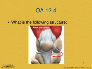
Knee Joint Anatomy • Tibiofemoral joint • condyles of the femur • __________________________________ • ______________________________________ • Tibial plateaus • flattened, very slightly concave • “Screw home mechanism” • _______________________________ • _____________________________________________________________________________
Knee Anatomy - Ligaments • 4 main ligaments- help stabilize knee jt • Medial Collateral (Tibial Collateral) • _______________________________________ • Attaches to medial femoral condyle and anterior medial tibia • Lateral Collateral (Fibular Collateral) • ________________________________ • Attaches to lateral femoral condyle and head of fibula
Knee Anatomy - Ligaments • Anterior Cruciate (ACL) • __________________________________________________________________ • attaches to lateral femoral condyle → medial tibia • Posterior Cruciate (PCL) • ______________________________________________________________________ • attaches to medial femoral condyle →lateral tibia
Knee Anatomy - Menisci • Menisci • firbrocartilage discs • Functions:1)____________________________________2) absorption and dissipation of force3) congruency of the surface to improve wt distribution4) • Thicker along the lateral portion
Menisci Cont • Poor blood supply (only outer 1/3 receives direct blood supply) • ____________________________________ • The medial is more commonly injured because of its attachment to the MCL ligament & more securely attached to the tibia (which makes it less mobile)
Knee Joint Anatomy • Patellofemoral joint • patella • ________________________________________________________________________ • femur • Patellofemoral groove • Q angle • The angle of pull of quadriceps on the patella • normal is 13 degrees male/ 18 female • How to measure?
Knee Anatomy - Muscles • Muscles-contribute to jt stability • Quadriceps (EXT): Vastus lateralis, vastus medialis, rectus femoris, vastus intermedius; quads also aid in patella alignment • Hamstrings (Flex): Semitendinosus (IR), Semimembranosus (IR), Biceps Femoris (ER) • Gastroc (Flex), Sartorius (Flex/IR), Gracilis (Flex/IR)
Prevention of Knee Injuries • Physical Conditioning and Rehabilitation • Total body conditioning is required • Strength, flexibility, cardiovascular and muscular endurance, agility, speed and balance • Muscles around joint must be conditioned (flexibility and strength) to maximize stability • Must avoid abnormal muscle action through flexibility • In an effort to prevent injury, extensibility of hamstrings, erector spinae, groin, quadriceps and gastrocnemius is important
Rehab (16-11), Functional Prophylactic Knee Braces (Fig 16-4) • Used to prevent and reduce severity of knee injuries • Provide degree of support to unstable knee • Can be custom molded and designed to control rotational forces and tibial translation
Patellofemoral- “Cho-Pat” strap (Fig 16-21) and horseshoe knee sleeve (Fig 16-20) • Used for several different conditions
Treatment of Knee Injuries • Normal acute protocol and NSAIDs • _______________________________ • Control swelling, fit for crutches if necessary,increase ROM and strength • Return to competition the safest and quickest way possible thru rehab, functional activities, and sports specific activities
Assessing the Knee Joint • Determining the mechanism of injury is critical • History- Current Injury • Past history • Mechanism- what position was your body in? • ____________________________ • Did you hear or feel anything? • Could you move your knee immediately after injury or was it locked? • _____________________ • Where was the pain
History - Recurrent or Chronic Injury • What is your major complaint? • When did you first notice the condition? • Is there recurrent swelling? • _______________________________ • _______________________________ • _______________________________ • Does it ever feel like giving way? • What does it feel like when ascending and descending stairs? • _________________________________
Observation • Walking, half squatting, going up and down stairs • Swelling, ecchymosis • Assessment of muscle symmetry/atrophy • What is the athlete’s level of function? • Does the athlete limp? • Full weight bearing? • Does athlete exhibit normal knee mechanics during function?
Palpation • Should assess bony structures checking for pain, bony deformity, warmth, swelling, spasms, and crepitus • Soft tissue • Lateral ligaments • _____________ • Assess for pain and tenderness • _____________ • _____________
Special Tests for Knee Instability • Laxity is a positive sign for injury • Translation (tibial translation) refers to the glide of tibial relative to the femoral • As the damage to stabilization structures increases, laxity and translation also increase • Valgus and Varus Stress Tests • Used to assess the integrity of the MCL and LCL respectively • Testing at 0 degrees incorporates capsular testing while testing at 30 degrees of flexion isolates the ligaments
Valgus Stress Test Varus Stress Test
Lachman Drawer Test • Will not force knee into painful flexion immediately after injury • Reduces hamstring involvement • At 30 degrees of flexion an attempt is made to translate the tibia anteriorly on the femur • A positive test indicates damage to the ACL
ACL, PCL and Meniscus Testing • Anterior Drawer- • Posterior Drawer- • McMurray’s Click-
Apley’s Compression Test • Hard downward pressure is applied w/ rotation • Pain indicates a meniscal injury • Used to detect meniscus tear
Recognition and Management of Specific Injuries • Medial Collateral Ligament Sprain • Cause of Injury • Result of severe blow or outward twist – valgus force • Signs of Injury - Grade I • Little fiber tearing or stretching • ________________________ • Little or no joint effusion • Some joint stiffness and point tenderness on lateral aspect • _________________________
Signs of Injury (Grade II) • Complete tear of deep capsular ligament and partial tear of superficial layer of MCL • No gross instability; __________________ • Slight swelling • ________________________________________ • Pain along medial aspect of knee • Signs of Injury (Grade III) • Complete tear of supporting ligaments • _____________________________________ • Minimum to moderate swelling • Immediate pain followed by ache • ____________________________________________________________________ • Positive valgus stress test
Care • RICE for at least 24 hours • Crutches if necessary • Knee immobilizer may be applied • Move from isometrics and STLR exercises to bicycle riding and isokinetics • Return to play when all areas have returned to normal • Continued bracing may be required
Care • Conservative non-operative approach for isolated grade 2 and 3 injuries • __________________________________________________________________________ • Follow with 2-3 week period of protection with functional hinge brace • When normal range, strength, power, flexibility, endurance and coordination are regained athlete can return • Some additional bracing and taping may be required
Lateral Collateral Ligament Sprain • Cause of Injury • _______________________________________________________________________ • Direct blow is rare** WHY? • Signs of Injury • Pain and tenderness over LCL • Swelling and effusion around the LCL • Joint laxity w/ varus testing • Care • Following management of MCL injuries depending on severity
Anterior Cruciate Ligament Sprain • Cause of Injury • MOI – athlete decelerates with foot planted and turns in the direction of the planted foot forcing tibia into internal rotation • May be linked to inability to decelerate valgus and rotational stresses - landing strategies • Male versus female
ACL – c/s cont • Research is quite extensive in regards to impact of femoral notch, ACL size and laxity, malalignments (Q-angle) faulty biomechanics • __________________________________________________________________________________ • ____________________________________________________________
Signs of Injury • Experience pop w/ severe pain and disability • Rapid swelling at the joint line • Positive anterior drawer and Lachman’s • Care • RICE; use of crutches • Arthroscopy may be necessary to determine extent of injury • Could lead to major instability in incidence of high performance • W/out surgery joint degeneration may result • Age and activity may factor into surgical option • Surgery may involve joint reconstruction w/ grafts (tendon), transplantation of external structures • Will require brief hospital stay and 3-5 weeks of a brace • Also requires 4-6 months of rehab
Posterior Cruciate Ligament Sprain • Cause of Injury • Most at risk during 90 degrees of flexion • Fall on bent knee is most common mechanism • _________________________ • Signs of Injury • Feel a pop in the back of the knee • Tenderness and relatively little swelling in the popliteal fossa • ________________________________________ • Care • RICE • Non-operative rehab of grade I and II injuries should __________________________ • Surgical versus non-operative • Surgery will require 6 weeks of immobilization in extension w/ full weight bearing on crutches • ROM after 6 weeks and PRE at 4 months
Meniscus Injuries • Cause of Injury • Medial meniscus is more commonly injured due to ligamentous attachments and decreased mobility • Also more prone to disruption through twisting and valgus forces • ________________________________________________________________________ • Signs of Injury • Diagnosis is difficult • Effusion developing over 48-72 hour period • Joint line pain and loss of motion • Intermittent locking and giving way • Pain w/ squatting
Care • Immediate care = PRICE • If the knee is not locked, but indications of a tear are present further diagnostic testing may be required • Treatment should follow that of MCL injury • If locking occurs, anesthesia may be necessary to unlock the joint w/ possible arthroscopic surgery follow-up • W/ surgery all efforts are made to preserve the meniscus -- with full healing being dependent on location • Torn meniscus may be repaired using sutures
Bursitis • Cause of Injury • Acute, chronic or recurrent swelling • _______________ = continued kneeling • _______________ = overuse of patellar tendon • Signs of Injury • Prepatellar bursitis may be localized swelling above knee that is ballotable • Presents with cardinal signs of inflammation • Swelling in popliteal fossa may indicate a Baker’s cyst • Care • _________________________________ • Aspiration and steroid injection if chronic
Iliotibial Band Friction Syndrome (Runner’s Knee) • Cause of Injury • Repetitive/overuse conditions attributed to mal-alignment and structural asymmetries • Can be the result of running on crowned roads • Signs of Injury • Irritation at band’s insertion • Tenderness, warmth, swelling, and redness over lateral femoral condyle • Pain with activity • Care • Correction of mal-alignments • Ice before and after activity, proper warm-up and stretching; NSAID’s • Avoidance of aggravating activities
Acute Patella Subluxation or Dislocation • Cause of Injury • Deceleration w/ simultaneous cutting in opposite direction (valgus force at knee) • _____________________________________ • ____________________________________ • ____________________________________ • More commonly seen in female athletes • Signs of Injury • W/ subluxation, pain and swelling, restricted ROM, palpable tenderness over adductor tubercle • Dislocations result in total loss of function • First time dislocation = assume fx
Care • Immobilize and refer to physician for reduction • Ice around the joint • Following reduction, immobilization for at least 4 weeks w/ use of crutches • After immobilization period, horseshoe pad w/ elastic wrap should be used to support patella • Muscle rehab focusing on muscle around the knee, thigh and hip are key (STLR’s are optimal for the knee)
Chondromalacia patella • Cause • Softening and deterioration of the articular cartilage • Possible abnormal patellar tracking due to genu valgum, foot pronation, patella alta, shallow femoral groove, increased Q angle, laxity of quad tendon • Signs of Injury • ______________________________________ • ________________________________________________________________________ • Care • Conservative measures • RICE, NSAID’s, isometrics for strengthening • Avoid aggravating activities • Surgical possibilities
Patellar Tendinitis (Jumper’s or Kicker’s Knee) • Cause of Injury • ________________________________________________________________________________________________________ • ______________________________________ • Signs of Injury • Pain and tenderness at inferior pole of patella and on posterior aspect of patella with activity • Care • Avoid aggravating activities • Ice, rest, NSAID’s • Exercise • Patellar tendon bracing • Transverse friction massage
Osgood-Schlatter Disease • Cause of Condition • _____________________________________ • __________________________________________________________________________________ • Resolves w/ aging • Signs of Condition • Both elicit swelling, hemorrhaging and gradual a bump occurring at the tibial tubercle • Pain with activity and tenderness over anterior proximal tibial tubercle
Care • Conservative • Reduce stressful activity until union occurs (6-12 months) • Padding may be necessary for protection • Possible casting, ice before and after activity • Isometerics