Download

1 / 42
430 likes | 2.21k Vues
The Immune Response: Antigen-Antibody Reactions. Introduction. Antibodies are bifunctional - they bind to the target antigen they recognize as foreign, and they enable other defense components to react with it Variable domain (Fab) - binds to target antigen

E N D
Antibodies are bifunctional - they bind to the target antigen they recognize as foreign, and they enable other defense components to react with it • Variable domain (Fab) - binds to target antigen • Constant domain (Fc) - interacts with cells of the immune system and other host defense mechanisms
occurs within the pocket formed by folding the VH and VL regions of the Fab domain
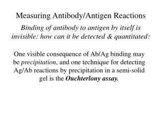
Binding is due to weak, noncovalent bonds • Shapes of epitope and binding site must be complementary for efficient binding • The high complementarity provides for the high specificity associated with antigen-antibody binding
The complement system is a series of protein components that must be activated in a cascade fashion (i.e., the activation of one component results in the activation of the next, and so on) • Results in lysis of antibody-coated bacteria and eukaryotic cells • Mediates inflammation • Attracts and activates phagocytic cells
There are three activation pathways • Each results in destruction of the target cell, but their triggering mechanisms are different • The classical pathway is dependent on antigen-antibody interactions to trigger it; it is fast and efficient
The lectin pathway is activated by mannose-binding lectins (MBLs) that have been secreted by liver; activation leads to opsonization • The alternative pathway does not require antigen-antibody binding; it is nonspecific and inefficient, but contributes to innate resistance
The final step in the pathway is the formation of a membrane attack complex that creates a pore in the membrane of the target cell • The pore allows entry of destructive enzymes or leads to osmotic rupture of the target cell
Ag-Ab Interactions – cont. • Toxin neutralization - antibody (antitoxin) binding to toxin renders the toxin incapable of attachment or entry into target cells • Viral neutralization - binding prevents viral attachment to target cells • Adherence inhibiting antibodies - sIgA prevents bacterial adherence to mucosal surfaces
Antibody-dependent cell-mediated cytotoxicity - involves the complement system, NK cells, or release of cytotoxic mediators from effector cells that attach to the target cell by means of Fc receptors • IgE and parasitic infection - in the presence of elevated IgE levels, eosinophils bind parasites and release lysosomal enzymes
Opsonization • Prepares the microorganism for phagocytosis • Phagocytes recognize the Fc portion of IgG or IgM antibodies coating the surface of the foreign microorganism • Phagocytosis can also be stimulated by components of the complement system, whether initiated by the classical or alternative pathways
Inflammation can be mediated by IgE attachment to mast cells and basophils, or by the binding of one of the complement components to mast cells and platelets • This complement component is also a powerful chemoattractant for macrophages, neutrophils, and basophils
Immune complex formation - two or more antigen-binding sites per antibody molecule lead to cross-linking, forming precipitins (molecular aggregates) or agglutinins (cellular aggregates) • Agglutination that specifically involves red blood cells is called hemagglutination • In vivo testing involves immediate or delayed skin testing for the presence of antibodies to various antigens
Agglutination - visible clumps or aggregates of cells or of coated latex microspheres • If red blood cells are agglutinated, this is called hemagglutination • Agglutination inhibition can be used to detect serum antibodies or to detect the presence of specific substances (e.g., illegal drugs) in urine samples by a competition assay
Box 33.2 Rapid urine testing for drugs, e.g cocaine – Abuscreen method
Figure 33.9 – Latex agglutination test for pregnancy Positive pregnancy test
Figure 33.10 – Viral Hemagglutination Some viruses bind to RBC and cause hemagglutination. If serum contains anti-viral Abs, then hemagglutination is inhibition – positive test.
Figure 33.11: Tube agglutination test for determining antibody titer.
Q: What is the titer in rows A-H? Figure 33.11: A microtiter plate illustrating hemagglutination. Ab in wells 1-10. Positive control = row 11, negative control = row 12. RBCs added to each well. If enough Ab is available to agglutinate the cells, they bind as a mat to the bottom of the well. If insufficient Ab is available, they form a pellet at the bottom.
Complement fixation • Irreversible alterations to complement components that are initiated by the binding of antibody to antigen; • Used to detect the presence of serum antibodies, thereby indicating prior exposure to a pathogen
If immune complexes are formed, then complement is used up and lysis will not occur when sensitive indicator cells are added • If immune complexes are not formed, then complement is not used up and lysis will occur when sensitive indicator cells are added
Enzyme-linked immunosorbent assay (ELISA) • Indirect immunosorbent assay - detects serum antibody • Antigen is coated on test wells and serum is added • If antibodies are present, they will bind antigen; if not, they will wash off
Add to the plate an enzyme that is covalently coupled to a second antibody against first immunoglobulin • If antigen was present, the second antibody will bind; if not, it will wash off • Add colorless substrate (chromogen) for the enzyme and measure colored product formation spectrophotometrically; • No colored product will form if everything washed off
Double antibody sandwich assay - detects antigens in a sample • Antibody is coated onto test wells and sample is added • If antigen is present in sample it will bind; if not it will wash off • React with antibody against the antigen; if antigen was present in the sample, this antibody will bind; if not, it will wash off • Continue with steps (c), (d), and (e) as in the indirect assay
Immunodiffusion - involves the precipitation of immune complexes in an agar gel after diffusion of one or both components • Single radial immunodiffusion (RID) assay - quantitative
Figure 33.15 Mancini technique – Single radial immunodiffusion assay
Double diffusion assay (Ouchterlony technique) - lines of precipitation form where antibodies and antigens have diffused and met; determines whether antigens share identical determinants
Figure 33.15 Ouchterlony technique – Double diffusion agar assay
Immunoelectrophoresis - antigens are first separated by electrophoresis according to charge, and are then visualized by the precipitation reaction • Greater resolution than diffusion assay
Figure 33.16 Classical Immunoelectrophoresis - Used to separate major blood proteins in serum for diagnostic tests Precipitin arcs form where Ab and Ag precipitate.
Immunofluorescence - dyes coupled to antibody molecules will fluoresce (emit visible light) when irradiated with ultraviolet light • Direct - used to detect antigen-bearing organisms fixed on a microscope slide • Indirect - used to detect the presence of serum antibodies
Immunoprecipitation - soluble antigens form insoluble immune complexes that can be detected by formation of a precipitin
Figure 33.18 – Immunoprecipitation – Precipitin curve and precipitation ring test.
Neutralization - an antibody that is mixed with a toxin or a virus will neutralize the effects of the toxin or the infectivity of the virus; this is determined by subsequent assay
Radioimmunoassay (RIA) - purified antigen labeled with a radioisotope competes with unlabeled sample for antibody binding • Serotyping - antigen-antibody specificity is used to differentiate among various strains (serovars) of an organism