Download
1 / 18
220 likes | 938 Vues
Introduction to Scanning Electron Microscopy @CMSE. What is an SEM? ____________________________________________________________. Scanning Electron Microscope is an instrument that investigates the surfaces of solid samples using a beam of electrons rather than light to form the image.

E N D
Introduction to Scanning Electron Microscopy @CMSE
What is an SEM? ____________________________________________________________ • Scanning Electron Microscope is an instrument that investigates the surfaces of solid samples using a beam of electrons rather than light to form the image.
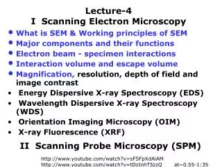
Advantages of using an SEM instead of a light microscope ____________________________________________________________ • Imaging • High depth of field • high resolution where features can be examined at high magnification. • Elemental Analysis - EDS • Structural Analysis - EBSD • Ease of sample preparation since most SEMs only require the sample to be conductive.
Advantages of using an SEM instead of a light microscope ____________________________________________________________ • There are a number of differences between light and electron optics. • light optics relies on the refraction or reflection of light. The wavelength of the visible light used in optical microscopes is between 400 and 700 nanometers (nm). Light microscopes are limited by the wavelength of imaging light to about 200 nanometers (0.2 microns). • In electron optical systems, electrostatic or magnetic fields are used to influence the trajectories of beams of electrons. Scanning electron microscopy uses electrons with energies of a few thousand electron volts, imaging a thousand times greater than that of visible light (2 to 3 eV). The wavelength is 0.02 nanometers.
What you observed? ____________________________________________________________ • Surface topography of metals, ceramics and minerals • Microstructural morphology of metals, ceramics and minerals
Imaging in SEM ____________________________________________________________ • High Magnification • Range ~ 5x to 500,000x • Resolution down to 1nm. point-to-point • High depth of field • Over 100x better than optical microscope • All specimen heights in focus simultaneously • Best at low magnification • High brightness guns - LaB6, FEG • Digital Storage • ESEM • Low keV, high resolution
SEM Operation Mode ____________________________________________________________ • High Resolution Mode: small beam, small current, optimum final aperture size after astigmatism correction, short working distance, high magnification • High-Current Mode: High beam current, better contrast and microanalysis (more electrons and x-rays are emitted and collected), larger aperture-poorer resolution, use at low magnification under 1000x so that image blur is not noticeable • Depth-of-Field Mode: small final aperture, long working distance, best at low magnification • Low-Voltage Mode: increased surface sensitivity, reduced specimen charging, beam size larger than at higher kv-poorer resolution
What kind of Data you get in SEM ____________________________________________________________ Topography (SE or GSE) • The surface features of an object or how it looks, its texture, detectable features limited to a few manometers. • These images give valuable information about sample's topography
What kind of Data you get in SEM ____________________________________________________________ Morphology (BSE) • The shape, size and arrangement of the particles making up the object that are lying on the surface of the sample or have been exposed by grinding or chemical etching, detectable features limited to several nanometers • This image gives valuable information about sample's composition Dr Anthony Garratt-Reed
What kind of Data you get in SEM ____________________________________________________________ Composition (EDS) • The elements and compounds the sample is composed of and their relative ratios, in areas ~ 1 micrometer in diameter • These X-Ray map images give valuable information about sample's composition SEM image Copper Tin Lead Dr Anthony Garrett-Reed
What kind of Data you get in SEM ____________________________________________________________ Crystallographic Information (EBSD) • The arrangement of atoms in the specimen and their degree of order; only useful on single-crystal particles >20 micrometers • These Orientation Imaging Micrographs give valuable information about sample's crystallographic orientation EDAX Inc.
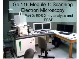
SEM Instruments Available in the EM Lab: _______________________________________________________ JEOL 5910: General-Purpose SEM Philips FEI XL30 : FEG-ESEM JEOL 6320: High-resolution SEM Secondary detectors (SE) Backscatter detectors (BSE) Energy-dispersive X-ray detectors (EDS)
JEOL 5910 General-Purpose SEM _______________________________________________________ • Large sample chamber • Easy to operate • Use a tungsten filament • Resolution down to 50 nm
FEI XL30 FEG-ESEM _________________________________________________________ • Large sample chamber with CCD camera • Use a field emission gun (FEG) • Gaseous secondary detector (GSE) used under poor vacuum in the environmental mode • Backscatter diffraction patterns (EBSD) • High resolution down to 20 nm
JEOL 6320 High-resolution SEM _________________________________________________________ • Restricted sample chamber • Use a field emission gun (FEG) • Higher resolution down to 1.4 nm
MT-X Ultra microtomeat Room Temperature ___________________________________________________________________________ • This microtome is used to obtain ultra-thin sections for electron microscopy, semi-thin and thick sections for light microscopy. • It can be used for both ultramicrotomy at room temperature sectioning and cryo-ultramicrotomy at low temperature sectioning
MT-X Ultra microtomecryo-ultramicrotomy at low temperature mode ___________________________________________________________________________ Cryo-ultramicrotomy at low temperature sectioning of polymers, elastomers, or any materials requiring processing down to -185°C. Each section of the sample can be between 5nm to 5um.
Introduction to Scanning Electron Microscopy Patrick Boisvert Thu Jan 24, 10-11:00am, 13-2137 The lecture will provide an introduction to the basic principles of Scanning Electron Microscopy with an approach to EDX, EBSD, and BSE. Contact: Patrick Boisvert, 13-1018, x3-3317, pboisver@mit.edu