Download

1 / 53
540 likes | 751 Vues
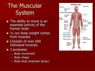
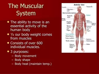
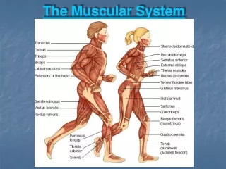
The Muscular System. Chapter 6. link. Muscle Tissues. Muscles make up nearly ½ of the body’s mass! Function: contraction/shortening movement! Muscle Types (Table 6.1, page 182) Skeletal Cardiac Smooth. Skeletal Muscle. “fiber” – elongated cells Striated, voluntary

E N D
The Muscular System Chapter 6 link
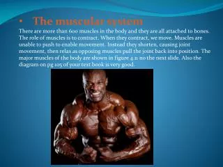
Muscle Tissues • Muscles make up nearly ½ of the body’s mass! • Function: contraction/shortening movement! • Muscle Types (Table 6.1, page 182) • Skeletal • Cardiac • Smooth
Skeletal Muscle • “fiber” – elongated cells • Striated, voluntary • Skeletal muscle is organ of the Muscular System • Surrounded by fascia separated into: • Endomysium: surrounds each fiber • Perimysium: surrounds groups of fibers (fascicle) • Epimysium: covers entire muscle • Continuous with tendons or aponeuroses
Skeletal Muscle • Figure 6.1, page 183
Anatomy of a Skeletal Muscle “Bundles of Bundles” Muscle (fascicles wrapped in epimysium) Fascicles (muscle fibers wrapped in perimysium) Fibers (elongated cells, enclosed with sarcolemma) Myofibrils (organelles which fill sarcoplasm) Myofilaments (threadlike protein filaments bundled into myofibrils)
Muscle Function • Producing movement • Muscles, bones, & joints work together • Maintaining posture & body position • Stabilizing joints • Generating heat (thermogenesis) • 80% of heat comes from muscle contraction (lost when bonds in ATP broken) • Shivering to raise temperature when cold
Microscopic Anatomy of Skeletal Muscle • Cells are multinucleate • Sarcolemma: cell membrane • Myofibrils: ribbon-like organelles that fill the cytoplasm • Alternating light (I) and dark (A) bands on myofibrils (striations) • Sarcomere: contractile unit of muscle (myofibrils) • Aligned end to end • Myofilaments within myofibrils • Sarcoplasmic reticulum (SR): specialized smooth ER • Stores calcium and releases it on demand when muscle fiber stimulated to contract
Myofilaments • Myosin filaments: thick filaments • Made of bundles of protein myosin • Contain ATPase enzymes (split ATP) • Extend length of A band • Ends have small projections called myosin heads (cross bridges): link thick & thin filaments together during contraction • Actin filaments: thin filaments • Made of contractile protein actin • Regulatory proteins prevent binding of myosin heads to actin • Anchored to the Z disc • I band only contains actin filaments
Properties of Skeletal Muscle • Excitability/responsiveness • Receive and respond to stimuli • Contractility • Ability to shorten (forcibly) when adequately stimulated • Extensibility • Muscle cells can be stretched • Elasticity • Ability to recoil and resume their resting length after being stretched
Neuromuscular Junction • Must be stimulated by nerve impulses • One motor neuron (nerve cell) can stimulate a few muscle cells or hundreds of them • Motor unit: one motor neuron and all skeletal muscle cells it stimulates • Axon of neuron branches into axon terminals, each of which forms junctions with sarcolemma of different muscle cells • Neuromuscular junctions: contain vesicles filled with neurotransmitters • NTM that stimulates skeletal muscle cells: acetylcholine (ACh) • Synaptic cleft: gap between axon terminal and sarcolemma of muscle cell; filled with interstitial fluid
Physiology of Muscle Contraction When nerve impulse reaches axon terminals… 1. Calcium channels open and Ca2+ enters the terminal causing release of Ach 2. ACh diffuses across the synaptic cleft and attaches to receptors (membrane proteins) that are located in the sarcolemma of the muscle cell 4. Ach stimulates sarcolemma and causes depolarization, generating an action potential (more on that in the nervous system!) 5. Action potential spreads throughout the muscle cell and stimulates sarcoplasmic reticulum to release calcium ions into the cytoplasm 6. Calcium ions trigger binding of myosin to actin, initiating filament sliding 7. Acetylcholinesterase enzyme breaks down ACh (present on sarcolemma and in synaptic cleft). • Single nerve impulse produces only one contraction • Prevents continued contraction of the muscle cell in the absence of additional nerve impulses.
Sliding Filament Theory • Pages 191-192 (fig 6.7 & 6.8) • Myosin heads attach to binding sites on the thin filaments when stimulated by nervous system (Calcium present) • Each cross bridge attaches and detaches (bend, break, & reform) several times during a contraction (using energy from ATP), generating tension that helps to pull the thin filaments toward the center of the sarcomere (form cross bridges further down actin filament) – power stroke • Myofilaments do not shorten – just slide past each other • Occurs simultaneously in sarcomeres throughout the muscle cell (cell shortens) • Attachment of myosin cross bridges to actin requires calcium ions • Takes a few thousandths of a second animation
Muscle Response • Threshold stimulus: minimal stimulus needed to cause contraction • All-or-none response • Can be measured by a myogram(figure 6.9)
Contraction of Skeletal Muscle as a Whole • Whole muscle contraction is through graded responses • different degrees of shortening throughout the whole muscle • Produced two ways: • Changing the frequency of muscle stimulation • Changing the number of muscle cells being stimulated at one time
Muscle Response to Increasingly Rapid Stimulation • Nerve impulses delivered to muscle at a very rapid rate (no chance for muscle to relax) • Effects of successive contractions are added together (summative) • Causes stronger & smoother contraction • Sustained contraction • Fused (complete) tetanus: contractions completely smooth and sustained • Unfused (incomplete) tetanus: up until the point of fused tetanus
Muscle Response to Stronger Stimuli • Force of muscle contraction mostly depends on how many cells are stimulated • when all motor units are active and all cells stimulated, muscle contraction is as strong as it can get • Muscle cells stimulated at the same time & entire muscle contracts • Motor units recruit other fibers Link
Energy for Muscle Contraction • Muscle store a small amount of ATP (a few seconds’ worth) to get going • ATP only energy source can be used directly to power muscle activity & must be regenerated continuously if contraction is to continue • ATP regeneration catalyzed by ATPase • Three pathways for ATP regeneration: • Direct phosphorylation of ADP by creatine phosphate • Aerobic respiration • Anaerobic glycolysis & lactic acid formation • Only 25% of energy is used – rest released as heat!
Creatine Phosphate • High energy molecule found in muscle fibers but not other cell types • Regenerates ATP in a fraction of a second by transferring a phosphate group to ADP • CP supplies exhausted in less than 15 seconds
Aerobic Respiration • 95% of ATP used for muscle activity • Oxidative phosphorylation: occurs in mitochondria, metabolic pathways that use oxygen • Glucose broken down completely to carbon dioxide & water, energy released as bonds are broken energy captured in ATP molecules • Generates a large amount of ATP • Slow and requires continuous delivery of oxygen and nutrient fuels to muscle to keep it going
Anaerobic Glycolysis & Lactic Acid Fermentation • If oxygen & glucose delivery is inadequate (intense muscle activity), glycolysis moves into lactic acid fermentation instead of oxidative phosphorylation • Only 5% as much ATP produced without oxygen • 2.5 times faster and provides most of ATP needed for 30-40 seconds of strenuous activity • Lactic acid accumulates and produces muscle soreness
Muscle Fatigue & Oxygen Deficit • Muscle fatigue: unable to contract even though being stimulated • Oxygen deficit occurs after prolonged muscle activity, causing fatigue • Work that a muscle can do and how long without fatigue depends on blood supply • Without adequate oxygen, lactic acid accumulates and ATP supply runs low, which leads to fatigue • i.e. marathon runners
Types of Muscle Contractions • Isotonic contractions: myofilamentsslide past each other, muscle shortens, movement occurs • Isometric contractions: muscles do not shorten, tension increases in muscle but myofilaments do not slide past each other • Movement pitted against an immoveable object • Muscle tone: continuous partial contractions that occur involuntarily due to nervous stimulation to keep a muscle firm, healthy, and ready for action • If no longer stimulated in this way, loses tone and muscle is flaccid and begins to atrophy
Muscle Movement • “Golden Rules” (Table 6.2, page 197) • All skeletal muscles cross at least one joint (with few exceptions) • Bulk of skeletal muscle lies proximal to joint crossed • All skeletal muscles have at least two attachments: the origin & the insertion • Skeletal muscles can only pull; they can never push • During contraction, a skeletal muscle insertion moves toward the origin
Muscle Movement • All skeletal muscles are attached to bone or to other connective tissue structures at no fewer than two points • Origin: attached to the immovable or less movable bone • Insertion: attached to the movable bone, and when muscle contracts, moves toward the origin • Some muscles have interchangeable origins & insertions or multiple origins/insertions (i.e. biceps brachii) • body movement occurs when muscles contract across joints
Biceps Brachii • Fig 6.12, page 197 • 2 origins at shoulder • Insertion across elbow – attaches to ulna Belly
Interactions of Skeletal Muscles • Muscles function in groups • Groups of muscles that produce opposite movements lie on opposite sides of a joint • Prime mover: muscle that has major responsibility for causing a movement • Antagonists: muscles that oppose or reverse a movement • Muscles can be both prime movers and antagonists (i.e. biceps & triceps) • Synergists: help prime movers by producing same movement or reducing undesirable movements (stabilize joints)
Putting it all together: Physics!!
Naming Skeletal Muscles • Direction of the muscle fibers • External obliques • Relative size of the muscle • Pectoralis major • Location of the muscle’s origin and insertion • Sternocleidomastoid • Shape of the muscle • deltoid • Action of the muscle • Extensor digitorum
Direction of Muscle Fibers • External oblique • oblique= at a slant • Rectus abdominus • Rectus = straight (parallel) • Using the midline of the body or axis of a long bone
Relative Size of Muscle • Major/minor • Vastus • Long; covers a lot of area; big • Maximus/minimus • Medius • Longus
Location • Frontalis • Temporalis • Tibialis anterior • Biceps femoris (not the same as the biceps brachii in your arm!)
Action of the Muscle • Adductor • Extensor digitorum
Number of Attachments • Biceps brachii • Biceps femoris • Triceps brachii Also give location (brachial; femoral)
Arrangement of Fascicles • Determines range of motion and power • Circular: concentric rings (sphincters) • Ex. Orbicularis muscles (eyes & mouth) • Convergent: fascicles converge toward a single insertion tendon (triangular or fan shaped) • Ex. Pectoralis major • Parallel: length of fascilces run parallel to the long axis of muscle (straplike) • Fusiform: spindle-shaped with expanded “belly” • Ex. Biceps brachii • Pennate: short fascicles attach obliquely to central tendon • Unipennate (insert into one side of tendon) • Bipennate & multipennate (most powerful)
Figure 6.15 Link
Circular • Orbicularis oculi • Orbicularis oris
Convergent • Pectoralis Major
Parallel • Sternocleidomastoid • Sartorius
Fusiform • Biceps brachii • Tensor fasciae lata • Semitendinosus
Unipennate • Extensor digitorumlongus