Download

1 / 19
210 likes | 402 Vues
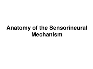
Anatomy of the Sensorineural Mechanism. The Bony Labyrinth. (from Zemlin, 1998). Three main subdivisions of the labyrinth cochlea semicircular canals (vestibular system) vestibule (vestibular system, mainly). Bony labyrinth : Hard, bony outer shell. (from Minifie, Hixon, & Williams, 1973).

E N D
The Bony Labyrinth (from Zemlin, 1998) • Three main subdivisions of the labyrinth • cochlea • semicircular canals (vestibular system) • vestibule (vestibular system, mainly)
Bony labyrinth: Hard, bony outer shell. (from Minifie, Hixon, & Williams, 1973) Membranous labyrinth: Fully contained inside the bony labyrinth; like a convoluted-shaped water balloon stuff inside the bony labyrinth.
Another view of the membranous labyrinth (from Perkins & Kent, 1986)
Again with the membranous labyrinth, this time all by itself; i.e., with the entire bony labyrinth stripped away. (from Deutsch & Richards, 1979)
Schematic and partially unrolled view of the middle ear and labyrinth showing the scala vestibuli, the scala media (also cochlear duct), and the scala tympani, along with the cochlear fluids (endolymph & perilymph). (adapted from Zemlin, 1998)
Cuts through an unrolled and a rolled cochlea (from Deutsch & Richards, 1979)
Cut through an unrolled cochlea (from Deutsch & Richards, 1979)
Detail of a single cut through the cochlea showing the Organ of Corti,8th N fibers entering through a tunnel in the spiral lamina (the habenula perforata), the spiral ligament, and the stria vascularis. (from Deutsch & Richards, 1979)
Notice the stria vascularis (also area vascularis) – the only good picture I have of this. The s.v. secretes endolymph. Notice also the spiral ligament, which attaches the b.m. to the bony wall of the cochlea, and the limbus(or limbus spiralis), a fibrous covering of the spiral lamina. (from Zemlin, Fig. 6-48) modiolus spiral ligament
Detail of the Organ of Corti (from Stevens,1951) Any cut through the cochlea will show 1 inner hair cell (IHC) and 3 (sometimes 4) outer hair cells (OHCs). This unit – 1 IHC and 3-4 OHCs is referred to as a hair cell channel. There are about 3000 channels in the human cochlea. (That number will become important later when we discuss cochlear implants.)
Electron micrograph of a hair bundle as seen from above. Note: (1) cilia are arranged according to height, (2) cilia are arranged in a very distinctive pattern, variously described as a ‘W’ or sometimes a ‘V’ shape. modiolus spiral ligament
IHCs OHCs spiral ligament modiolus
The cochlear and vestibular braches of the 8th cranial nerve (from Deutsch & Richards, 1979)