Download
1 / 12
120 likes | 450 Vues
Dislocation of the DSEK Donor Graft into the Posterior Segment An Intraoperative Complication in DSEK Surgery. Mark M Fernandez MD, Mark S Gorovoy MD, George OD Rosenwasser MD, Terry Kim MD, Alan N Carlson, MD, and Natalie A Afshari MD Financial Disclosures:

E N D
Dislocation of the DSEK Donor Graft into the Posterior SegmentAn Intraoperative Complication in DSEK Surgery Mark M Fernandez MD, Mark S Gorovoy MD, George OD Rosenwasser MD, Terry Kim MD, Alan N Carlson, MD, and Natalie A Afshari MD Financial Disclosures: Dr. Gorovoy has a relationship with Harvey Instruments Dr. Rosenwasser is a paid lecturer for Allergan, Vistacon and Inspire Dr. Afshari has a research grant from Reseach to Prevent Blindness Drs. Fernandez, Kim and Carlson report no financial interests
Introduction • Descemet’s stripping endothelial keratoplasty (DSEK) is rapidly becoming the preferred treatment for corneal edema due to endothelial dysfunction • DSEK surgery is also being conducted in eyes with prior vitrectomy • We present a series of DSEK graft dislocations into the posterior segment Anterior chamber OCT showing a well adhered DSEK graft
Methods • Four cases of intraoperative DSEK graft dislocation into the posterior segment were identified • The surgical management, and the final outcome of each eye are discussed A DSEK graft dislocated posteriorly
Case 1: • A 59 year old woman with a sutured sulcus intraocular lens and history of pars plana vitrectomy underwent DSEK surgery for decompensation of her fourth full thickness corneal graft • As the donor graft unfolded within the anterior chamber, it slipped through a hole in the posterior capsule and entered the posterior segment • Attempts to float the graft using irrigation were unsuccessful, so incisions were closed • Seven days later she underwent pars plana vitrectomy to remove the graft from the posterior segment as well as full thickness penetrating keratoplasty
Case 2: • A 62 year old man with Fuchs dystrophy underwent DSEK surgery three months after phacoemulsification with anterior vitrectomy and sulcus intraocular lens placement • The donor graft was inserted into the anterior chamber and unfolded uneventfully; filtered air was inserted into the anterior chamber and the DSEK graft could no longer be visualized • A superotemporal sclerotomy was made by a vitreoretinal surgeon; the donor graft was seen laying on the surface of the macula and was removed. It was then repositioned in the anterior chamber • The graft remained attached postoperatively, but it did not clear. Two months later, repeat DSEK surgery was performed
Case 2: Two months after repeat DSEK, the new donor graft is well-adhered. A healed scleral incision is seen superotemporally
Case 3: • A 63 year old man with a failed penetrating keratoplasty, aphakia, only remnants of an iris rim, and prior pars plana vitrectomy underwent secondary scleral sutured posterior intraocular lens placement, followed by a DSEK one month later • During DSEK surgery, the graft dislocated posteriorly around the sutured posterior chamber intraocular lens during unfolding with the irrigator/aspirator (see supplemental video) • Using the I&A handpiece, the graft was maneuvered around the sutured posterior chamber IOL into the anterior chamber and repositioned • Post-op day one revealed a dislocated donor graft. The patient underwent a penetrating keratoplasty two weeks later
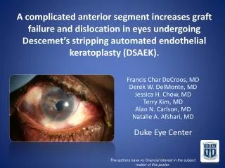
Case 4: • A 79 year old woman with history of traumatic open globe, pars plana vitrectomy, trabeculectomy, Ahmed valve, sutured posterior chamber lens and a failed large diameter corneal graft underwent DSEK surgery • Upon opening, the globe began to collapse. Air was used to maintain the anterior chamber. The graft was inserted and the globe was refilled with balanced salt solution • Air was injected to unfurl the graft. Despite attempts to hold it in the anterior chamber, the graft slipped between the lens implant and ciliary body into the posterior segment • A pars plana vitrectomy was performed nine days later to remove the graft. Two months after, a penetrating keratoplasty was performed
The gray-colored donor graft is held in place centrally with a Sinskey hook while air is inserted into the anterior chamber The graft is seen within the anterior chamber moments before it slips into the posterior segment Shortly after, the graft has dislocated into the posterior segment and is no longer visible
Results: Patient and Intraoperative Risk Factors • All eyes had undergone anterior or pars plana vitrectomy between three months and several years prior to surgery • All eyes had undergone complicated intraocular lens placement prior to DSEK surgery; All IOLs were in the posterior chamber. Three were sutured and one was a sulcus IOL • In two cases, the graft dislocated as it unfolded within the anterior chamber • In two cases, dislocation occurred after injection of sterile air within the anterior chamber
Results: Surgical Management • Of the four cases reported, one eye underwent successful repeat DSEK and the other three underwent successful PK following graft removal • In two cases the misplaced graft was subsequently repositioned and remained at least partially attached. Both grafts failed and were repeated within two months • In one case the graft was retrieved using the I&A handpiece. In three cases a vitreoretinal surgeon was consulted to remove the graft • Satisfactory visual results were attained after the complication was addressed in each case
Conclusions • Dislocation of donor grafts to the posterior segment is a rare complication of DSEK surgery that appears to require repeat corneal grafting, however good final results can be obtained • This complication must be considered in the preoperative planning of DSEK surgery in vitrectomized eyes, especially those with other risk factors for posterior dislocation like aphakia, pseudophakia with open posterior capsule, and sutured IOL • Although access to a vitreoretinal surgeon for the removal of posterior segment grafts is ideal, in some circumstances the problem can be addressed using tools available for anterior segment surgery