Download
1 / 1
10 likes | 335 Vues
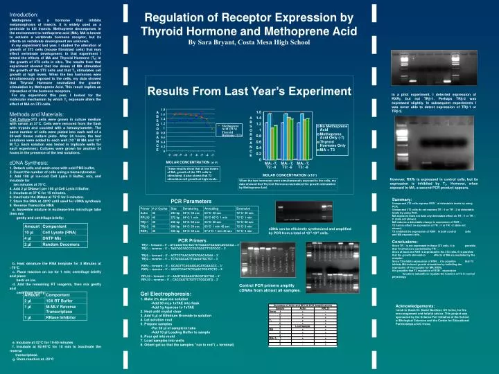
ABSORBANCE MOLAR CONCENTRATION (x10 x ) Results From Last Year’s Experiment These results show that at low doses of MA, growth of the 3T3 cells is stimulated. It also shows that T3 stimulates cell growth at high levels.

E N D
ABSORBANCE MOLAR CONCENTRATION (x10x) Results From Last Year’s Experiment These results show that at low doses of MA, growth of the 3T3 cells is stimulated. It also shows that T3 stimulates cell growth at high levels. When the two hormones were simultaneously exposed to the cells, my data showed that Thyroid Hormone neutralized the growth-stimulation by Methoprene Acid. PCR Parameters Primer # of Cycles Size Denaturing Annealing Extension Actin 40 294 bp 94°C/ 30 sec 60°C/ 30 sec 72°C/ 30 sec RPL30 40 272 bp 94°C/ 1 min 58°C-60°C/ 1 min 72°C/ 1 min TR-1 40 250 bp 94°C/ 60 sec 55°C/ 60 sec 72°C/ 60 sec TR-2 40 600 bp 94°C/ 50 sec 65°C/ 1 min 45 sec 72°C/ 3 min cDNA can be efficiently synthesized and amplified by PCR from a total of 10-5-10-6 cells. RXR 40 350 bp 94°C/ 50 sec 67.5°C/ 1 min 45 sec 72°C/ 3 min PCR Primers TR1 - forward – 5’ – ATCAGGTGCTACTCTGAAGTGAGGCAGGCGA – 3’ TR1 – reverse – 5’ – TAGTGGTACCCTGTGGCTTTGTCCC – 3’ TR2 – forward – 5’ – ACTCCTAACAGTATGACAGAA – 3’ TR2 – reverse – 5’ – TCTGGGCACTTGAGATGCTCT – 3’ RXR – forward – 5’ – GCAGTTCAGAGGACATCAAGCC – 3’ RXR – reverse – 5’ – GCCCTCACTCTCAGCTCGCTCTC – 3’ RPL30 – forward – 5’ – AAGTGGGAAGTACGTGCTGG – 3’ RPL30 – reverse – 5’ – CACCAGTCTGTTCTGGCATG – 3’ * * * Control PCR primers amplify cDNAs from almost all samples. Gel Electrophoresis: 1. Make 2% Agarose solution -Add 50 mLs 1xTAE into flask -Add 1g Agarose to 1xTAE 2. Heat until crystal clear 3. Add 5 µl of Ethidium Bromide to solution 4. Let solution cool 5. Prepare samples -Put 50 µl of sample in tube -Add 10 µl Loading Buffer to sample 6. Pour gel into mold 7. Load samples into wells 8. Orient gel so that the samples "run to red"( + terminal) Regulation of Receptor Expression by Thyroid Hormone and Methoprene Acid By Sara Bryant, Costa Mesa High School Introduction: Methoprene is a hormone that inhibits metamorphosis of insects. It is widely used as a pesticide to kill insects. Methoprene decomposes in the environment to methoprene acid (MA). MA is known to activate a vertebrate hormone receptor, but its effects on vertebrate development are unknown. In my experiment last year, I studied the alteration of growth of 3T3 cells (mouse fibroblast cells) that may effect vertebrate development. In that experiment I tested the effects of MA and Thyroid Hormone (T3) in the growth of 3T3 cells in vitro. The results from that experiment showed that low doses of MA stimulated the growth of the 3T3 cells and that T3 stimulates cell growth at high levels. When the two hormones were simultaneously exposed to the cells, my data showed that Thyroid Hormone neutralized the growth-stimulation by Methoprene Acid. This result implies an interaction of the hormone receptors. For my experiment this year, I looked for the molecular mechanism by which T3 exposure alters the effect of MA on 3T3 cells. Methods and Materials: Cell Culture-3T3 cells were grown in culture medium with serum at 37°C. Cells were removed from the flask with trypsin and counted with a hemacytometer. The same number of cells were plated into each well of a 24-well tissue culture plate. After 24 hours, the test solutions were added to each well (10-7 M MA and 10-4 M T3). Each solution was tested in triplicate wells for each experiment. Cultures were grown for another 24 hours in the presence of the test solutions. cDNA Synthesis: 1. Detach cells and wash once with cold PBS buffer. 2. Count the number of cells using a hemacytometer. 3. Add 100 µl ice-cold Cell Lysis II Buffer, mix, and incubate for ten minutes at 75°C. 4. Add 2 µl DNase I per 100 µl Cell Lysis II Buffer. 5. Incubate at 37°C for 15 minutes. 6. Inactivate the DNase at 75°C for 5 minutes. 7. Store the RNA at -20oC until used for cDNA synthesis 8. Reverse Transcribe RNA a. Assemble mixture in nuclease-free microfuge tube then mix gently and centrifuge briefly: b. Heat denature the RNA template for 3 Minutes at ~70°C c. Place reaction on ice for 1 min; centrifuge briefly and place back on ice. d. Add the remaining RT reagents, then mix gently and centrifuge briefly: e. Incubate at 42°C for 15-60 minutes f. Incubate at 92-95°C for 10 min to inactivate the reverse transcriptase. g. Store reaction at -20°C In a pilot experiment, I detected expression of RXR, but not TR-1. Perhaps TR-2 was expressed slightly. In subsequent experiments I was never able to detect expression of TR-1 or TR-2. However, RXR is expressed in control cells, but its expression is inhibited by T3. However, when exposed to MA, a second PCR product appears. Summary: Unexposed 3T3 cells express RXR at detectable levels by using PCR. Unexposed 3T3 cells do not express TR-1 or TR-2 at detectable levels by using PCR. MA exposure does not have any detectable effect on TR-1 or TR-2. (data not shown). MA induces a detectable change in expression of RXR. T3 had no effect on expression of TR-1 or TR-2 (data not shown). T3 inhibited the expression of RXR in both control cells and MA exposed cells. Conclusions: Since TR is not expressed in these 3T3 cells, it is possible that T3 effects are controlled by TR. Since at least one RXR is expressed in the 3T3 cells, it is possible that the growth stimulation affects of MA are mediated by the receptor. Since T3 inhibits expression of RXR, it is possible that T3 inhibits MA-induced growth stimulation by inhibiting the expression of the receptor for MA (RXR). It is possible that T3 regulation of RXR expression functions naturally to regulate the function of T3 in normal physiology. Acknowledgements: I wish to thank Dr. David Gardiner, UC Irvine, for his encouragement and helpful advice. This project was sponsored by the Science Fair Initiative of the School of Biological Sciences and the Center for Educational Partnerships at UC Irvine.


