Download
1 / 74
780 likes | 1.04k Vues
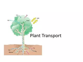
Plant Transport. Plants. Plant: terrestrial (mostly), multicellular, photoautotrophic, eukaryote, true tissues and organs. Plant Structure . Tissue Basic Tissue Types: pg 717 - give rise to specialized cells Dermal - outer coat Vascular – transport tubes

E N D
Plants Plant: terrestrial (mostly), multicellular, photoautotrophic, eukaryote, true tissues and organs
Plant Structure Tissue Basic Tissue Types: pg 717 - give rise to specialized cells • Dermal - outer coat • Vascular – transport tubes • Ground – between Dermal and Vascular
Basic Tissue Layout Dermal Ground Vascular
Dermal tissue Ground tissue Vascular tissue
Dermal Tissue • Epidermis • Function: protection: secretes the cuticle, forms prickles and root hairs
Thorns, Spines and Prickles Based on where they originate • Thorns – modified stems • Spines – modified leaves • Prickles – modified epidermal cells
Prickle Rose “thorns” are prickles “A rose between two prickles.”
Vascular Tissue Xylem: Water conducting – unidirectional (up) • Dead at maturity – pg 719 Phloem: Sugar conduction – bidirectional • Sieve Tube Members: alive and functional – lack many organelles • Companion Cells: connected to Sieve Tube Members by plasmodesmata – supports the STM with its organelle function
WATER-CONDUCTING CELLS OF THE XYLEM SUGAR-CONDUCTING CELLS OF THE PHLOEM Sieve-tube members: longitudinal view Tracheids 100 m Vessel Companion cell Pits Sieve-tube member Sieve plate Tracheids and vessels Nucleus Vessel element Vessel elements with partially perforated end walls 30 m 15 m Tracheids Companion cell Cytoplasm Figure. 35.9
Ground Tissue • Occupies the space between the vascular tissue and the dermal tissue • Functions: • Storage – roots and stems • Support – stems • Photosynthesis – leaves and some stems
Types of Ground Tissue 1.Parenchyma: undifferentiated, thin cell walls (still flexible) – used for metabolism and photosynthesis Ex: Pallisade and Spongy Mesophyll of leaf Potato, Fruit pulp 2. Collenchyma: unevenly thickened cell walls – support young parts of plants – no lignin, but stronger than parenchyma Ex: “Strings” in celery 3. Sclerenchyma: highly thickened cell walls – lignified – support mature tissue – hard and dead Two types: Fibers and Sclerids Ex: Walnut Shell, Stone Cells in Pears
SCLERENCHYMA CELLS PARENCHYMA CELLS COLLENCHYMA CELLS 5 m 80 m Cortical parenchyma cells Sclereid cellsin pear 25 m Cell wall Parenchyma cells 60 m Collenchyma cells Fiber cells Figure 35.9
Plant Parts: Roots, Stems and Leaves Roots: Functions: - absorb water, nutrients and minerals - anchor plant in soil - store food and water - support the plant
(a) Prop roots (b) Storage roots (c) “Strangling” aerialroots (d) Buttress roots (e) Pneumatophores
Increasing Absorption • Root hairs – extensions of the epidermis • Branching roots – lateral roots • Mycorrhizae
Root Structure • Outside In • Epidermis (D) • Cortex (G) – storage and nutrient transfer • Endodermis (G) – separates ground and vascular tissue – important for water transfer • Pericycle (V) – forms the lateral roots • Stele (Xylem and Phloem) (V)
Epidermis Cortex Vascular cylinder Endodermis Pericycle Core of parenchyma cells Xylem Phloem 100 m 100 m (a) Transverse section of a typical root. In the roots of typical gymnosperms and eudicots, as well as some monocots, the stele is a vascular cylinder consisting of a lobed core of xylem with phloem between the lobes. (b) Transverse section of a root with parenchyma in the center. The stele of many monocot roots is a vascular cylinder with a core of parenchyma surrounded by a ring of alternating xylem and phloem. Endodermis Key Dermal Pericycle Ground Vascular Xylem Phloem 50 m
Eudicot Root – Cross Section • From: http://www.inclinehs.org/smb/Sungirls/images/dicot_stem.JPG
Monocot Root Cross Section • From: http://www.inclinehs.org/smb/Sungirls/images/monocot_stem.JPG
Monocot Stele • From: http://www.botany.hawaii.edu/faculty/webb/BOT201/Angiosperm/MagnoliophytaLab99/SmilaxRotMaturePhloemXylem300Lab.jpg
100 m Emerging lateral root 2 1 4 3 Cortex Vascular cylinder Epidermis Lateral root Growth of Lateral Roots
Eudicot – tap root Monocot – fibrous roots Eudicot & Monocot Roots - External
Stems Function: - support leaves and flowers - photosynthesis (non-woody plants – herbaceous) - storage: food (tubers – potato) and water (cactus)
Stem Structure • Nodes: points where leaves are/were attached • Internodes: area of growth between the nodes • Bud: Developing leaves • Terminal/Apical Bud: end of a branch • Lateral/Axillary Bud: lateral growth – between leaf petiole (“stem” of leaf) and main stem • Bud Scale Scars: Sites of old bud scales (protective layers around the buds) - # of bud scale scars indicates the age of the stem • Leaf Scars: Sites where leaves were attached to the stem • Lenticles: “bumps” of cork lined pores that allow for oxygen exchange in the stem
Terminal bud Bud scale Axillary buds Leaf scar This year’s growth (one year old) Node Stem Internode One-year-old side branch formed from axillary bud near shoot apex Leaf scar Last year’s growth (two years old) Scars left by terminal bud scales of previous winters Leaf scar Growth of two years ago (three years old)
Stem: Internal Anatomy Epidermis Ground Tissue Pith Vascular Bundles Contain Xylem and Phloem May contain: Vascular Cambium, Cork Cambium, Sclerenchyma
Groundtissue Epidermis Vascularbundles 1 mm (b) A monocot stem. A monocot stem (maize) with vascularbundles scattered throughout the ground tissue. In such anarrangement, ground tissue is not partitioned into pith andcortex. (LM of transverse section) Monocot Stem Structure
Monocot Stem Vascular Bundles Xylem Phleom
Monocot Stem Vascular Bundle • From: http://iweb.tntech.edu/mcaprio/stem_dicot_400X_cs_E.jpg
Phloem Xylem Sclerenchyma(fiber cells) Ground tissueconnecting pith to cortex Pith Key Cortex Epidermis Dermal Vascularbundle Ground Vascular 1 mm (a) A eudicot stem. A eudicot stem (sunflower), withvascular bundles forming a ring. Ground tissue towardthe inside is called pith, and ground tissue toward theoutside is called cortex. (LM of transverse section) Eudicot Stem Structure
Eudicot Stem Cross Section • From: http://plantphys.info/plant_physiology/images/stemcs.jpg
Eudicot Stem Vascular Bundle Sclerenchyma Phloem Xylem Vascular Cambium
Leaves Functions: - photosynthesis - storage (succulent leaves, Aloe) - protection: spines, toxins, trichomes - reproduction: flowers (modified leaves)
Leaves Functions: - photosynthesis - storage (succulent leaves, Aloe) - protection: spines, toxins, trichomes - reproduction: flowers (modified leaves)
Leaves: External Structure • Blade • Petiole • Stipule • Axillary Bud • Veins
Leaves: Internal Structure - Cuticle - Upper Epidermis (Adaxil) - Mesophyll:- Palisade Layer - Spongy Layer - Air Spaces - Vascular Bundle - Bundle Sheath Cells - Xylem and Phloem -Lower Epidermis (Abaxil) - Stomata - Guard Cells - Cuticle
Guard cells Key to labels Dermal Stomatal pore Ground Vascular Epidermal cell Sclerenchyma fibers 50 µm Cuticle (b) Surface view of a spiderwort (Tradescantia) leaf (LM) Stoma Upper epidermis Palisade mesophyll Bundle- sheath cell Spongy mesophyll Lower epidermis Guard cells Cuticle Vein Xylem Vein Air spaces Guard cells Guard cells Phloem Figure 35.17a–c 100 µm Transverse section of a lilac (Syringa) leaf (LM) (c) (a) Cutaway drawing of leaf tissues
Turgor loss in plants causes wilting • Which can be reversed when the plant is watered Figure 36.7
EXTRACELLULAR FLUID CYTOPLASM – + H+ + – ATP H+ – + H+ Proton pump generates membrane potential and H+ gradient. H+ H+ H+ – H+ + H+ – + Plant Transport of Solutes • Proton Pumps: Active transport of H+ out of the cell • Builds proton gradient • Functions: provides potential for the COTRANSPORT of materials across the membrane with the H+
+ – Plant cells can also accumulate a neutral solute, such as sucrose ( ), by cotransporting down the steep proton gradient. + H+ – H+ NO3 – + – H+ H+ H+ NO3– S Cell accumulates anions (, for example) by coupling their transport to theinward diffusion + – + – H+ H+ H+ – H+ + H+ H+ NO3– S H+ H+ H+ H+ – S H+ of through a cotransporter. H+ H+ NO3– H+ – NO3 – + NO3 – S S – S – H+ + + H+ NO3– – – H+ + H+ S H+ + H+ H+ + – (c) Contransport of a neutral solute (b) Cotransport of anions Figure 36.4c Figure 36.4b