Download
1 / 51
520 likes | 651 Vues
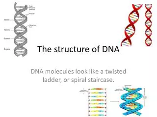
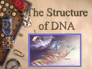
The Structure of DNA. This is a very well known story But done here! Using 1B physics. Real importance still emerging. Please read my source. Contemporary Physics, volume 44, number 4, July – August 2003, pages 289 – 305. Outline. The People The Problem (DNA)

E N D
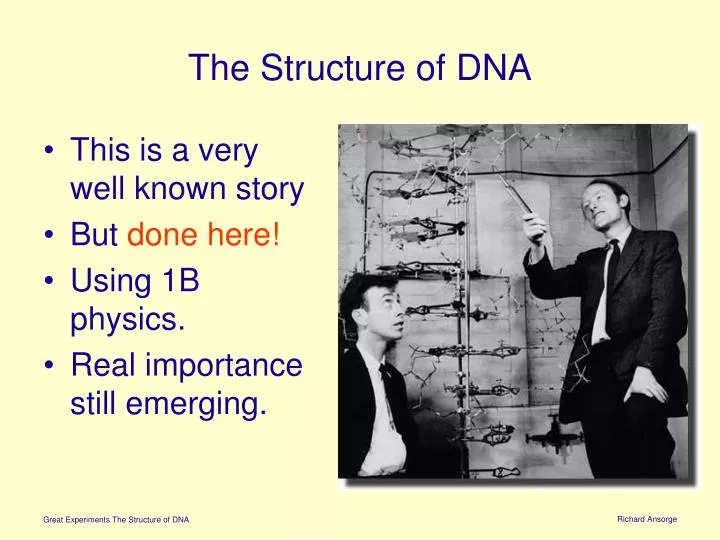
The Structure of DNA • This is a very well known story • But done here! • Using 1B physics. • Real importance still emerging. Richard Ansorge
Please read my source Contemporary Physics, volume 44, number 4, July – August 2003, pages 289 – 305 Richard Ansorge
Outline • The People • The Problem (DNA) • X-ray diffraction patterns • Events in 1952/3 • Comments Richard Ansorge
Francis Crick Age 37 in 1953 Francis Crick was born on 8 June 1916 near Northampton. His father and uncle ran a boot and shoe factory. Soon after he was born his mother instructed her younger sister to carry him to the top of the house to ensure that in later life he would ‘rise to the top’. No doubt his subsequent career confirmed her in the efficacy of this treatment. His secondary schooling was first at Northampton Grammar School, and then at Mill Hill School in north London. In 1934 he entered University College, London and graduated 3 years later with a second-class Honours Degree in Physics with subsidiary Mathematics. He said later that he lost his religious faith at the age of 12, and his growing attachment to science was in part related to a desire to remove the ‘unfortunate vestiges of earlier beliefs’. In later life he resigned his Fellowship at Churchill College when the College decided to build a chapel. Richard Ansorge
James Watson Age 25 in 1953 James Watson was born in Chicago on 6 April 1928. He attended school in the Chicago public system. He proved very bright and was one of the original Quiz Kids in a wartime radio programme. He entered the University of Chicago at the age of 15 and graduated in 1947, specializing in zoology. He started graduate work at the University of Indiana under the supervision of Salvador Luria who, with his friend Max Delbrück, were leaders in the field of bacterial viruses. In May 1951, he attended a meeting at Naples, and heard Maurice Wilkins talking about his X-ray measurements on deoxyribonucleic acid (DNA). By chance, at a small meeting in the USA, Luria met John Kendrew, a colleague of Perutz, who was looking for someone to help with his investigation on the structure of the protein myoglobin. The matter was arranged, and Watson joined the Cavendish Laboratory in the autumn of 1951. Richard Ansorge
Rosalind Franklin Age 35 in 1953 Rosalind Franklin was born in London on 25 July 1920. Her father was a banker, and her family, Jewish uppermiddle class were in comfortable circum-stances. She was a clever child, and, after attending a private primary school, she was enrolled, at the age of 11, at St Paul’s School for Girls, a private school with a high academic reputation. In 1938 she entered Newnham College, Cambridge, and took the Natural Sciences Tripos, starting with physics, chemistry, mineralogy and mathematics in her first year, and ending with chemistry in Part II of the Tripos in her third and final year. She obtained an upper-second class in that examination, but obtained first classes in the two previous years. In 1950 she was awarded a 3-year Fellowship to work at a biophysics unit, headed by John Randall, at King’s College, London. In January 1951 King’s College had received some good specimens of DNA from Rudolf Signer, a Swiss biochemist at Berne; so Randall changed her programme to the X-ray study of the structures of these fibres. Richard Ansorge
Maurice Wilkins Age 37 in 1953 Maurice Wilkins was born in Pongaroa, New Zealand (of Anglo-Irish parents) on 15 December 1916. He attended St Edwards School, Birmingham, and in 1935 he entered St John’s College, Cambridge. He took the Natural Sciences Tripos and graduated in 1938, having obtained a lower-second class in Part II physics of the Tripos. He went to work under Randall at the University of Birmingham on luminescence in solids, a subject of considerable interest at the time for its application to radar screens. For this work he received a PhD in 1940. He was then sent to the University of California, where he worked on the separation of uranium isotopes, part of the Manhattan Project. Joined Randall’s the Biophysics Unit in King’s College in 1948, received DNA samples in 1950 and he started work on the X-ray investigation. His Naples talk aroused Watson’s interest in 1951. Richard Ansorge
Lawrence Bragg Age 63 in 1953 “The two Braggs had shared the 1915 Nobel Prize for Physics for inventing X-ray crystallography; Lawrence, at 25, was the youngest winner ever. His pride, however, became tinged with regret since it was assumed too easily by others that his father was the originator who was generously recognizing help from his son. In fact it was Lawrence - a research student at the time - who had the big idea about reflection from crystal planes, and who was later to show such skill in interpreting the diffraction patterns. Sir William's principal contribution lay in instrument development. Theirs was a real collaboration, but not without tensions. We youngsters who knew him as Cavendish Professor in post-war Cambridge knew nothing of the bouts of depression, the sense of inadequacy and the rare but explosive angers of the suave and kindly Edwardian gentleman who had charge of our destinies.” “Light is a Messenger: The Life and Science of William Lawrence Bragg”, Graeme K Hunter, 2004 OUP. Richard Ansorge
DNA Richard Ansorge
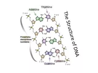
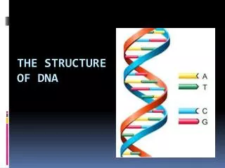
The chemical formula of DNA The DNA molecule is a very long chain polymer, the monomers of which are called nucleotides. Each nucleotide consists of three units: a negatively charged phosphate, a sugar known as deoxyribose, and a base. The nucleotides are linked along the phosphate – sugar units, which form the backbone of the molecule. A base is attached to each sugar. Richard Ansorge
NB Atomic Arrangement The atomic arrangement of a phosphate – sugar unit is shown here. All the units are identical. The atoms in the sugar unit are conventionally numbered as shown. The deoxy part of the name of DNA refers to the fact that there are two hydrogen atoms attached to the 2’ carbon atom. It should be noted that the atoms in the phosphate – sugar unit are not arranged in a plane. The four oxygen atoms in the phosphate unit are at the corners of a tetrahedron, with the phosphorus atom at its centre. The four carbon atoms and the oxygen atom in the sugar ring are also not in a plane but have a puckered arrangement. repeat repeat Richard Ansorge
Bases There are four bases: Thymine, Cytosine, Adenine and Guanine. ( A C G T) The sequence of the bases along the phosphate – sugar chain is irregular. It is this sequence that contains the genetic information in DNA. The chemical formula of nucleic acids was established mainly by Alex Todd and co-workers. Richard Ansorge
DNA Dimer of the deoxribonucleotide monomers adenine and guanine drawn such that A is to the left of and above G. Richard Ansorge
X-ray diffraction Richard Ansorge
X-Ray Diffraction • You have done this! • Diffraction pattern is FT of “aperture” • gaussian gaussian • top hat sinc • point wide • array of points array of points Richard Ansorge
FT of Helix? • Important problem in early 1950’s • In 1950, Pauling proposed the -helix structure for proteins (subsequent Nobel) • Crick and others had published in 1951 haemoglobin Richard Ansorge
Layers with FT of Helix diameter 2a This in fact is just the diffraction grating condition. The amplitude along the nth layer is proportional to: z pitch p x y where Jn is nthorder Bessel function Richard Ansorge
Pattern Shows that the diffraction pattern has cylindrical symmetry. Bessel functions of integral order look like damped sinusoidal functions. The X-ray pattern therefore contains ‘streaks’ located around the turning points of the Bessel functions. Richard Ansorge
Thus and Helix Parameters The properties of Bessel functions show that the first turning point occurs when: As a point on the helix moves through one pitch, z changes by P, and its projection on the x – y plane travels round a circle of circumference 2a. Thus the rise angle is given by Richard Ansorge
destructive interference Richard Ansorge
Two forms of DNA In October 1951 Franklin and Gosling found that there are two different X-ray patterns of DNA depending on the relative humidity of the sample. They correspond to different tructures, which they termed A and B. The A form occurs at a relative humidity of 75%, and the B form at a relative humidity of 90%. We may anticipate at this stage to say that both forms are double helices with slightly different dimensions. The A form is crystalline, while the B form exists as fibres. The two forms are readily interchanged by varying the relative humidity. G. L. Squires, Contemporary Physics, 44 (4), July – August 2003, pages 289 – 305 Richard Ansorge
Franklin’s X-ray Images Franklin decided to study the A form first Richard Ansorge
Building Models On 21 November 1951, Watson attended a colloquium at King’s College at which Franklin gave an account of her previous 6 months’ work on the A form of DNA. The next day he met Crick at Paddington for a journey to see Dorothy Hodgkin at Oxford and on the train gave him an account of Franklin’s talk. Crick was somewhat concerned that Watson had not made notes at the talk. However, there was no lack of confidence on their part, and on the basis of a probable helix structure, plus the experimental data of Franklin and Wilkins, they started to concoct a model. Unfortunately Watson, who at that stage knew little crystallography, had misunderstood what he had heard at the seminar. Franklin had reported that there were eight water molecules per nucleotide, and Watson mistakenly thought that this referred to the whole chain in a unit cell. So they underestimated the water content by a factor of 11. G. L. Squires, Contemporary Physics, 44 (4), July – August 2003, pages 289 – 305 Richard Ansorge
First Model In the next few days they built a model from pieces of metal lying around the Cavendish Laboratory, which had been used by Bragg, Kendrew and Perutz 18 months previously in an unsuccessful attempt to obtain the helical protein structure. The model consisted of a helix with three chains, with the sugar – phosphate chains on the inside and the bases on the outside. The three chains were held together by divalent cations of magnesium that were attached to the negatively charged phosphate groups. The model was completed by 27 November and Wilkins was invited to come and inspect it. G. L. Squires, Contemporary Physics, 44 (4), July – August 2003, pages 289 – 305 Richard Ansorge
Visit He arrived at the Cavendish Laboratory the next day, accompanied by Franklin, Gosling and William Seeds, another worker at King’s College. It did not take Franklin long to demolish the model. Firstly, she did not think that the X-ray data yet proved that the structure was helical, secondly, there was no room in the model for the correct amount of water, thirdly, she was of the opinion on chemical grounds that the phosphates would be on the outside and, finally, if there were divalent cations present, they would be surrounded by tight shells of water molecules and could not be centres of a tight structure. Watson records that the King’s College party took the 10.10 train from London in the morning and returned on the 3.40 from Cambridge the same day. As the visit included lunch, it can be seen that little time was wasted in the discussion. G. L. Squires, Contemporary Physics, 44 (4), July – August 2003, pages 289 – 305 Richard Ansorge
Fiasco The news of the fiasco soon reached Bragg. Bragg was not too pleased with Crick even before this episode. Crick was constantly criticizing the attempts made at the Cavendish Laboratory to elucidate the structure of proteins. He had given a seminar showing that, of the various methods, only one, namely isomorphous replacement, a technique for determining the phases of the diffraction spectra, had any chance of success. (He was subsequently proved right.) He had entitled the seminar What Mad Pursuit a quotation from Keats’ Ode to a Grecian Urn; he used it again for the title of his autobiography. Needless to say Bragg did not take kindly to this frank criticism from a research student. A little after the seminar when Crick was sitting behind Bragg just before the start of a lecture and voicing his views, probably in his usual loud voice, Bragg turned and said ‘Crick, you’re rocking the boat.’ On another occasion, Bragg complained that Crick’s voice made his head buzz. Randall and Bragg discussed the strategy for DNA and decided that future work was to be left to King’s College. Crick and Watson were told to stop working on DNA and to get back to what they were supposed to be doing. G. L. Squires, Contemporary Physics, 44 (4), July – August 2003, pages 289 – 305 Richard Ansorge
Competition The stimulus for a resumption of model building by the Cavendish pair was provided by Linus Pauling at the California Institute of Technology in the autumn of 1952 Pauling turned his attention to DNA. A colleague, Robert Corey, provided him with some diffraction pictures of DNA, and he proceeded to build a model structure. On 31 December, he and Corey sent a manuscript announcing their result to the Proceedings of the National Academy of Sciences, USA, and on the same day Linus wrote about the work to his son Peter, who was at the time a research student in the Cavendish Laboratory and was in fact sharing an office with Watson and Crick. When Watson heard Peter’s news, he was alarmed that Pauling with his formidable reputation had entered the quest for the structure of DNA. He asked Peter to write for a copy of his father’s manuscript, which reached him near the end of January 1953. G. L. Squires, Contemporary Physics, 44 (4), July – August 2003, pages 289 – 305 Richard Ansorge
Competition Pauling’s structure, like the previous model of Watson and Crick, was helical, with three chains in the helix, and the sugar – phosphate units on the inside. The three chains were held together by hydrogen atoms that neutralized the charges on the phosphate groups. Watson saw that this was a chemical blunder. Although momentarily heartened by this knowledge, he still worried that Pauling would soon see his mistake and try again. On 30 January, Watson went to London to visit Bill Hayes, a microbiologist at Hammersmith Hospital with whom he was collaborating on bacterial reproduction. On the way to Hammersmith Hospital he stopped at King’s College, and Wilkins showed him, in Franklin’s absence, her X-ray photograph of the B form of DNA. The picture was much better than any Watson had previously seen and confirmed his view that the structure was helical. G. L. Squires, Contemporary Physics, 44 (4), July – August 2003, pages 289 – 305 Richard Ansorge
Second attempt The next day back in Cambridge he went to Bragg to request that he and Crick could try model building again. He found Bragg in a receptive frame of mind. Pauling had already sent him a copy of his manuscript. Bragg had been very upset when Pauling succeeded in solving the a-helical protein structure, after his own incorrect attempt. He did not want to be beaten by his rival again. He therefore agreed that Watson and Crick could resume their model building, and the Cavendish Laboratory workshop was asked to make representations of the various atoms in DNA. It took a little time for the parts to arrive and they started with cardboard cutouts. Their aim was a model of the B form. They had two chains in the helix and at first continued with the sugar – phosphate backbones at the centre. However, they found that all the structures compatible with the X-ray data were unsatisfactory stereochemically, that is, some atoms were too close together, and after a few days changed to models with the backbones on the outside. G. L. Squires, Contemporary Physics, 44 (4), July – August 2003, pages 289 – 305 Richard Ansorge
Bases? On 19 February, Watson produced a model in which he paired identical bases between the two backbone chains; that is, he paired adenine with adenine, thymine with thymine, and so on. This scheme has the drawback that, since thymine (T) and cytosine (C) have single rings, while adenine (A) and guanine (G) have double rings, T –T and C–C pairs have smaller dimensions than A–A and G–G pairs. So the two backbones would have to be variable distances apart to accommodate the different pairs. Watson himself realized this but thought that the difficulty would not be insuperable. Donohue pointed out that Watson was using the wrong tautomeric forms of the bases. He should have used the keto form, which Donohue said was the common one, and not the enol form. These are illustrated in figure 11 for thymine. Watson told him that the textbooks that he had consulted gave the enol form, but Donohue stuck to his guns and said the textbooks were wrong. A consequence of using the keto form was that the differences between the T –T and C–C pairs and the A–A and G–G pairs were much larger than before. G. L. Squires, Contemporary Physics, 44 (4), July – August 2003, pages 289 – 305 Richard Ansorge
Final Model This was a setback, but Watson continued to play with his cardboard cutouts for the bases (the workshop had still not produced the metal representations) and on the morning of Saturday,28 February, he tried pairing adenine with thymine, and cytosine with guanine, using the keto forms of the bases. He saw that this structure met all the objections to his previous version. Pairing a base with a single ring with one with a double ring means that an A–T pair and a C–G pair have the same dimensions. When Crick came to the Cavendish Laboratory later in the morning, they realized that they had at last solved the problem. They repaired to their usual haunt, the Eagle pub in Bene’t Street, for lunch to tell everyone who would listen that they ‘had found the secret of life’. Crick went home and told his wife that they seemed to have made a big discovery. Years later she told him that she had not believed a word of it. ‘You were always coming home and saying things like that,’ she said. G. L. Squires, Contemporary Physics, 44 (4), July – August 2003, pages 289 – 305 Richard Ansorge
Richard Ansorge Reconstructed model in Science Museum
Afterwards • Bragg decided DNA was too BIG for the Cavendish • Set-up Molecular Biology Richard Ansorge
Subsequent Events Crick and Watson finished building the model of their structure on 7 March. Bragg, Perutz and Kendrew were shown the model, with enthusiastic commentaries by Crick. Todd was brought in to see it and pronounced it plausible biochemically. An elderly physicist, G. F. C. Searle, also came to see it. He had collaborated with J. J. Thomson in 1890 measuring the ratio of the electrostatic and electromagnetic units, and had dominated the teaching of practical physics in the Cavendish—not always with the approval of the rest of the staff—for over fifty years. When he saw the model, Searle, then aged eighty eight, announced that if this was the basis of human heredity ‘no wonder we’re such a queer lot’. He could have been told that it was the basis of heredity for all life. Wilkins came to Cambridge on 12 March and a few days later wrote to Crick ‘I think you are a couple of old rogues but you may well have something. I like the idea.’ Franklin came soon after and also accepted the model. Franklin tragically died of cancer on 16 April 1958 at the age of thirty seven. In 1962 Crick, Watson and Wilkins were awarded the Nobel prize in physiology or medicine for their work in elucidating the structure of DNA. Richard Ansorge
Other Evidence From 1945 to 1949, Erwin Chargaff and co-workers at Columbia University conducted a series of experiments in which they analysed the proportions of the four bases in the DNA taken from a wide variety of living organisms. A selection of their results is shown in Table 2. It can be seen that the amounts of adenine and thymine are essentially equal, and similarly for guanine and cytosine; the differences are within the experimental errors. The pairing suggested by Watson and Crick completely explains the Chargaff results. Richard Ansorge
Life – Very Complicated Richard Ansorge
DNA is the result of Evolution Richard Ansorge
Computing plays a vital role Richard Ansorge
Conclusions • Luck helps • Luck favours a prepared mind • The text books might be wrong • Simple physical models might be a short cut to deeper understanding • Teamwork? Richard Ansorge
After word Richard Ansorge
Cavendish Research Students and Staff 1952 Watson Crick Bragg Richard Ansorge
Computer Laboratory Staff ~1949 Richard Ansorge
The Old Computer Laboratory Richard Ansorge
Computer Laboratory: EDSAC first UK computer 1949 Richard Ansorge
COMPUTING DNA Scientific agenda for 21st Century? The Site in 2005 Richard Ansorge