Download
1 / 36
360 likes | 511 Vues
Complete course available at www.emsonline.net. Overview of CBT 434 Cardiovascular Emergencies. Introduction. Cardiovascular Emergencies Leading cause of death in the US Hundreds of thousands of Americans die from cardiac arrest each year

E N D
Complete course available at www.emsonline.net Overview of CBT 434 Cardiovascular Emergencies
Introduction • Cardiovascular Emergencies • Leading cause of death in the US • Hundreds of thousands of Americans die from cardiac arrest each year • 9 out of 10 cardiac arrest victims die before they get to the hospital
Practical Skills To receive CBT or OTEP credit, you must perform the following practical skills: • Focused history using SAMPLE/OPQRST • Assisting with nitroglycerin • Auscultation and assessing breath sounds • Assisting ventilations with BVM • Care for ACS, CHF, aortic dissection, shock • Use of AED
Terms acute coronary syndrome (ACS) - A term used to describe a range of symptoms and conditions from acute myocardial infarction to unstable angina aortic dissection- A tear in the lining of the aorta infarction - Death of tissue due to loss of blood flow ischemia - Poor oxygen supply to tissue myocardium - Another term for heart muscle
Terms, continued pulmonary edema - Abnormal accumulation of fluid in the tissues and air spaces of the lungs sustained tachycardia - Persistent heart rate of 100 or greater caused by a clinical condition such as hypoxia or impending shock thrombus - A clot formed in a blood vessel or in a chamber of the heart
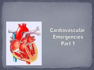
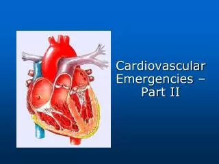
Anatomy • Neck to lower ribs • Divided into mediastinum and pleural cavities • Diaphragm at its base • Contains trachea, esophagus, heart, aorta, vena cava and the pulmonary artery • Pleural cavities Thoracic Cavity
Anatomy, continued • 12 pairs of ribs • Connect to sternum through a bridge of cartilage • Lower 5 ribs connect to sternum through the costal arch • Intercostal muscles between ribs Structures
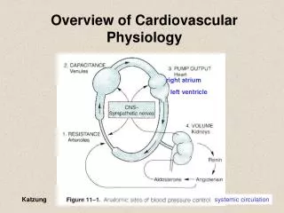
Anatomy, continued Arteries/Veins • Purpose of CV system • Provide cells with oxygen nutrients • Remove waste Components • Heart • Arteries • Arterioles • Capillaries • Veins • Venules
Anatomy, continued Coronary Arteries 1. Right coronary artery 2. Left coronary artery 3. Atria 4. Ventricles 1 2 3 4
Myocardial Ischemia Myocardial ischemia is the lack of blood flow and oxygen to the myocardium (heart muscle). • Inadequate blood flow to a part of the body • Caused by constriction or blockage of the blood vessels • Cells do not receive adequate supply of O2 occluded 75% occluded Coronary Artery
Myocardial Ischemia, continued • Obstruction • Inadequate blood flow to a part of the body • Caused by constriction or blockage of the blood vessels • Cells do not receive an adequate supply of oxygen
Myocardial Ischemia, continued • Reduced blood flow associated with conditions that cause: • Hypotension (e.g. blood loss) • Tachycardia • Bradycardia
Coronary Thrombosis 1 Plaque forms on the inner wall of an artery
Coronary Thrombosis, continued 1 Hard surface of the plaque tears, exposing the soft under side 2
Coronary Thrombosis, continued 1 2 Platelets arrive to form a blood clot 3
Other Sources of Chest Pain • Mediastinum • Chest wall • Lungs and pleura • Abdomen • Psychogenic Not all chest pain is cardiac related.
Conditions • Angina pectoris is chest pain due to myocardial ischemia. • Brought on by exercise, stress or cold weather • Possible radiation of pain to jaw, arm or upper back • Sudden onset • Usually relieved with rest within 3-5 minutes and/or nitro
Conditions, continued • Acute coronary syndrome (ACS) describes a range of clinical conditions. • The symptoms, which vary from patient to patient, are caused by acute myocardial ischemia: • Shortness of breath • Discomfort • Chest pain • Pressure • Nausea • Weakness • Dysrhythmia • Syncope
Conditions, continued Acute myocardial infarction (AMI) • Chest discomfort • Discomfort in other areas of the upper body • Shortness of breath • Diaphoresis, nausea or weakness
Conditions, continued • Aortic dissection • Blood gets behind inner layer of the aorta • Blood starts to fill space between layers of arterial wall • Aorta widens and significantly disrupts blood flow • Symptoms • Sudden and severe chest or upper back pain • Anxiety • Diaphoresis • Nausea
Conditions, continued • Congestive heart failure (CHF) • Occurs when heart is too weak to adequately circulate blood • In left-sided heart failure, pulmonary edema occurs as blood backs up into lungs • Increases fluid in alveoli - results in SOB
Conditions, continued Congestive heart failure signs • Fatigue • Cough, dyspnea • Pulmonary edema (a severe form of CHF) • Tachypnea • Agitation and confusion • Hypertension • Swollen feet or lower legs
Initial Assessment • Guides initial treatment • Quickly assess a patient • Make a decision SICK or NOT SICK
SICK/NOT SICK A SICK patient is one who can die quickly unless you initiate aggressive BLS and ALS treatment and rapid transport. A NOT SICK patient is one who can be ill or injured, but not severely enough to be life threatening.
Key Clinical Indicators • Respirations (rate, character) • Pulse (rate, character) • Mental status • Skin signs and color • Body position SICK or NOT SICK
Physical Exam • Auscultate breath sounds starting at the bases • BP in both arms (note difference of 10 mm Hg or more) • Skin color, moisture and temperature • Pulse oximetry • Blood glucometry • Head, neck-to-toe exam
Physical Exam, continued Atypical presentations are common in elderly, diabetics and females.
Patient Care • General steps • Decide SICK or NOT SICK • Ensure adequate airway and respirations • Administer oxygen • Position appropriately • Assure ALS response
Patient Care, continued • Other considerations • Control airway secretions • Assist ventilations with BVM • Prepare for cardiac arrest and application of an AED
Nitroglycerin • Used to treat angina • Relaxes vascular muscles • Increases blood flow & oxygen to myocardium
Nitroglycerin, continued • You may assist a patient in taking prescribed nitroglycerin if: • Pain is the same for which nitroglycerin is normally taken • 2. Patient’s BP is greater than 100 mmHg systolic *Follow your local protocol if different.
Nitroglycerin, continued • “Assisting” a patient with nitroglycerin means you can: • Locate the container • Open it • Offer a pill to the patient
Summary • Thoracic cavity is divided into mediastinum and pleural cavities. Structures within the thoracic cavity include: • Intercostal muscles • Ribs • Sternum • Costal arch • Diaphragm • Heart • Lungs • Trachea • Aorta • Pulmonary arteries
Summary, continued Myocardial ischemia is the lack of blood flow and oxygen to the heart muscle. Acute coronary syndrome (ACS) is the term used to describe clinical conditions ranging from unstable angina to acute myocardial infarction. Sources of chest pain include the mediastinum, chest wall, lungs/pleura and abdomen. It can also be due to psychogenic sources. Common cardiovascular emergencies are angina, AMI, aortic dissection, CHF and cardiogenic shock.
Summary, continued OPQRST is a mnemonic that helps assess the character of a complaint. Principles of care for a cardiovascular emergency include: • Decision of SICK or NOT SICK • Ensure an adequate airway and respirations • Administer oxygen • Position the patient appropriately • Assure an ALS response