Download
1 / 12
160 likes | 541 Vues
Pseudomonas and related organisms. Aerobic nonfermenters Pseudomonas aeruginosa : opportunistic infections of multiple sites Burkholderia cepacia : RT infection in cystic fibrosis patients, UTI, opportunistic infections Burkholderia pseudomallei : opportunistic pulmonary infections

E N D
Pseudomonas and related organisms Aerobic nonfermenters Pseudomonas aeruginosa: opportunistic infections of multiple sites Burkholderia cepacia: RT infection in cystic fibrosis patients, UTI, opportunistic infections Burkholderia pseudomallei: opportunistic pulmonary infections Stenotrophomonas maltophilia: opportunistic infections Acinetobacter baumannii:opportunistic infections of RT Moraxella catarrhalis: opportunistic RT infections
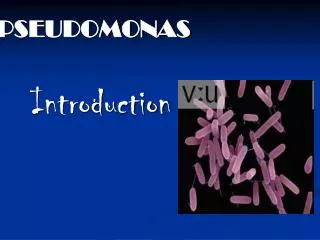
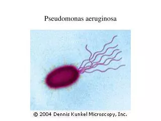
Pseudomonas Structure and Physiology Gram-negative rods. Motile with polar flagella. Obligate aerobe. Oxidase-positive. Do not ferment carbohydrates. Resistant to multiple drugs.
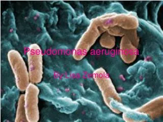
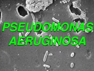
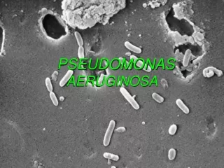
P. aeruginosa Forms round colonies with a fluorescent greenish color, sweet odor, and b-hemolysis. Pyocyanin- nonfluorescent bluish pigment; pyoverdin- fluorescent greenish pigment; pyorubin, andpyomelanin Some strains have a polysaccharide capsule. Identification of P. aeruginosa is usually based on colonial morphology, b-hemolysis, oxidase positivity, the presence of characteristic pigments and sweet odor, and growth at 42 oC.
P. aeruginosa Pathogenesis and Immunity This organism is widely distributed in nature and is commonly present in moist environments in hospitals. It is pathogenic only when introduced into areas devoid of normal defenses, e.g., 1. Disruption of mucous membrane and skin. 2. Usage of intravenous or urinary catheters. 3. Neutropenia (as in cancer therapy). P. aeruginosa can infect almost any external site or organ. P. aeruginosa is invasive and toxigenic. It attaches to and colonizes the mucous membrane or skin, invade locally, and produces systemic diseases and septicemia. P. aeruginosa is resistant to many antibiotics. It becomes dominant when more susceptible bacteria of the normal flora are suppressed.
P. aeruginosa Pathogenesis and Immunity Elastases: helps bacteria spread and inhibit neutrophil chemotaxis; induces antibodies in chronic infections. Hemolysins Phospholipase C; Rhamnolipid, also inhibits ciliary activity. Exotoxin A: causes tissue necrosis and is lethal for animals (blocks protein synthesis); immunosuppressive. Exoenzyme S and T: cytotoxic to host cells. Antigenic structure, enzymes, and toxins Pili and nonpilus adhesins. Polysaccharide capsules (alginate, glycocalyx): seen in cultures from patients with cystic fibrosis. LPS- endotoxin, multiple immunotypes. Pyocyanin: catalyzes production of toxic forms of oxygen that cause tissue damage.
P. aeruginosa Clinical Diseases Ear infections Otitis externa: mild in swimmers; malignant (invasive) in diabetic patients. Chronic otitis media Endocarditisseen in intravenous drug abusers. Urinary tract infection Sepsis: most cases originate from infections of lower RT, UT, and skin and soft tissue. *Ecthyma gangrenosum in sepsis: hemorrhagic necrosis of skin, often do not contain pus. Infection of wounds and burns (blue-green pus). *Verdoglobin or fluorescent pigment detected in wounds, burns, or urine by UV fluorescence. Skin and nail infections Meningitis (when introduced by lumbar puncture). Pulmonary infection Necrotizing pneumonia in CF patients (diffuse, bilateral bronchopneumonia with microabscess and necrosis). Eye infections: corneal ulcer.
P. aeruginosa Laboratory Diagnosis Specimen: skin lesions, pus, urine, blood, spinal fluid, sputum. Culture: blood agar plate and differential media. Identification of P. aeruginosa is described above. Several subtyping methods, including phage typing and molecular typing, are available for epidemiologic purposes. Treatment Combined antibiotic therapy is generally required to avoid resistance that develops rapidly when single drugs are employed. Avoid using inappropriate broad-spectrum antibiotics, which can suppress the normal flora and permit overgrowth of resistant pseudomonads. Injection of hyperimmune globulin and granulocyte transfusion to augment compromised immune function for selected patients.
P. aeruginosa Prevention and Control Pseudomonas spp. normally inhabit soil, water, and vegetation and can be isolated from the skin, throat, and stool of healthy persons. Spread is from patient to patient via contact with fomites or by ingestion of contaminated food and water. Methods for control of infection are similar to those for other nosocomial pathogens. Special attention should be paid to sinks, water baths, showers, hot tubs, and other wet areas. High risk population: patients with leukemia, burns, cystic fibrosis, and immunosuppression.
P. aeruginosa Prevention and Control Control: 1. Patients at high risk should not be admitted to a ward where cases of pseudomonas infection are present. 2. Patients infected with P. aeruginosa should be isolated. 3. Sterilize all instruments, apparatus, and dressing; antimicrobial and other therapeutic substances. 4. Monitor clinically relevant isolates of P. aeruginosa by a suitable typing system to identify epidemic strains.
Stenotrophomonas maltophilia A common nonfermentative, gram-negative isolate. It infects debilitated or immunocompromised persons, and causes a wide spectrum of diseases, including wound infections, UT infections, pneumonia, sepsis, meningitis, etc. It is resistant to many commonly used antibiotics, and patients receiving long-term antibiotic therapy are particularly at risk for acquiring infections. Infections may be acquired from contaminated disinfectants, respiratory therapy and monitoring equipment, and ice machines.
Burkholderia They colonize the moist environmental surfaces and are commonly associated with nosocomial infections. B. cepacia and B. pseudomallei are important pathogens. B. cepacia causes RT infections particularly in cystic fibrosis patients, UT infections and septicemia. Usually non-fatal except for RT infections in CF patients. B. pseudomallei usually causes opportunistic infections, but may sometimes infect previously healthy persons. Infection by this organism may result in asymptomatic infection, acute suppurative cutaneous infection that may progress to sepsis, and chronic pulmonary infection ranging in severity from mild bronchitis to necrotizing pneumonia.
2005/7/30 台南高雄疑似發生類鼻疽疫情,疾病管制局提醒民眾,皮膚如有傷口,請勿接觸污染的土壤或水源 疾病管制局今天公佈今年自七月11日至29日以來,類鼻疽累計通報16例,其中高雄縣9例、台南市4例、高雄市2例、台南縣1例。其中6例死亡,3例在加護病房,另7例住普通病房。類鼻疽係由類鼻疽伯克氏菌Burkholderia pseudomallei所造成的臨床感染症,屬假單孢菌屬革蘭氏陰性桿菌,此菌在土壤、水池及積水環境中存在,會感染馬、羊、豬等動物以及人類。其流行地域為東南亞地區及澳洲北部的熱帶地域。該局自89年即將此病納入監測。89年通報病例1例、90年15例、91年9例、92年5例、93年13例。本次疫情發生原因,疾病管制局初步調查研判可能係因日前南部豪大雨,將土壤中之病菌沖刷出來,所造成的民眾感染事件,病例多發生在二仁溪流域。該局鄭重呼籲在二仁溪流域附近居民,若有發燒等症狀者,務必迅速就醫。並告訴醫師居住地區,疾病管制局呼籲,醫師對於上述地區發燒病患,應先排除感染此病的可能性,若有懷疑應立即以抗生素治療,並採檢送驗。