Download
1 / 59

E N D
1. ULCERATIVE LESIONS OF THE ORAL CAVITY SAM J. CUNNINGHAM, MD,PhD
FRANCIS B. QUINN, JR., MD,FACS
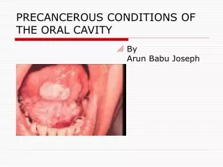
2. ORAL CAVITY LIPS
TEETH
GINGIVA
ORAL MUCOUS MEMBRANES
PALATE
TONGUE
ORAL LYMPHOID TISSUES
3. Acute: small, recent onset, short duration, recurrent Trauma
Recurrent Aphthous Stomatitis
Behcet�s
Herpesvirus Infection
Herpangina
4. Trauma: Cheek Biting
5. Trauma: Ill-Fitting dentures
6. Trauma: Chemical Burns
7. Trauma: Abrasions from Teeth
8. Recurrent Aphthous Stomatitis(RAS) Most common ulcerative lesion of oral cavity
Recurrent, painful ulcers
Confined to soft mucosa
Subdivided into three types:
Minor aphthae
Major aphthae
Herpetiform aphthae
9. Recurrent Aphthous Stomatitis(RAS) Minor aphthae:
Less than 1 cm
Heal completely in 7-10 days without scarring
Painful
Prodromal stage
Shallow and round to oval
Gray to yellow membrane
Clusters of up to 5 ulcers
Steroids
10. Recurrent Aphthous Stomatitis (RAS) Minor apthae
11. Recurrent Aphthous Stomatitis (RAS) Major Aphthae
Uncommon
Irregular, deep ulcers
1-3 cm in size
Raised borders
Heal in 4-6 weeks
Extensive scarring and distortion
BIOPSY!!
Steroids
12. Recurrent Aphthous Stomatitis (RAS) Major apthae
13. Recurrent Aphthous Stomatitis (RAS) Herpetiform Aphthae
Uncommon
Crops of up to 150 very small (<3mm) ulcers
Heal completely in 7-10 days
COMPLETELY UNRELATED TO HERPESVIRUS
14. Recurrent Aphthous Stomatitis (RAS) Herpetiform aphthae
15. Behcet�s Symptom complex of:
Recurrent aphthous ulcers of the mouth
Painful genital ulcers
Uveitis or conjuctivitis
16. Behcet�s Affects persons of Mediterranean, Middle Eastern, or Japanese decent
Easily confused with Stevens-Johnson syndrome or Reiter�s disease
Need referral for systemic treatment
17. Behcet�s
18. Herpesvirus Infection HSV-1 and/or HSV-2
Primary Infection
Secondary Infection
Varicella zoster virus (HHV-3)
19. Herpesvirus Infection Primary Infection
Herpetic gingivostomatitis
Younger patients
Often asymptomatic
May be associated with fever, chills, malaise
Vesicles-ulcers-crusting
Anywhere in the oral cavity
20. Herpesvirus Infection Primary Infection
21. Herpesvirus Infection Primary Infection
22. Herpesvirus Infection Secondary Infection
Reactivation of latent virus
Not associated with systemic symptoms
Small vesicles
Occur only on the hard palate and gingiva
Prodromal signs
23. Herpesvirus Infection Secondary infection
24. Herpesvirus Infection Varicella zoster virus (HHV-3)
Latent infection
Oral ulcers
Dermatomal distribution
25. Herpesvirus Infection Varicella zoster virus
26. Herpesvirus Infection Varicella zoster virus
27. Herpangina NOT caused by Herpesvirus
Coxsackie A virus
Children < 10 years of age
Common in summer and fall
Often subclinical presentation
Headache/Abdominal pain 48hrs prior to papulovesicular lesions on tonsils and uvula.
Sore throat
28. Herpangina
29. Chronic: longer duration, well circumscribed, raised borders, indurated base with crater Trauma
Infection
Neoplasm
Necrotizing sialometaplasia
30. Trauma: Ill-Fitting dentures
31. Infection Rare
HIV/AIDS patients
Bacterial
Deep mycotic infection
Candida
32. Infection Bacterial
Usually secondary infection
Primary infection: syphilis, tuberculous, or actinomycosis
33. Infection Bacterial-Syphilis
34. Infection Bacterial-Syphilis
35. Infection Mycotic
Blastomycosis
Histoplasmosis
36. Infection Histoplasmosis
37. Infection Candida
Candida albicans
Most common
Normal flora
Predisposing factors
White creamy patches
KOH prep
Nystatin oral suspension
38. Infection Candida
39. Neoplasm Squamous cell carcinoma (SCC)
Most common
Irregular ulcers with raised margins
May be exophytic, infiltrative or verrucoid
Mimic benign lesions grossly
40. Neoplasm Squamous cell carcinoma
41. Neoplasm Squamous cell carcinoma
42. Neoplasm Squamous cell carcinoma
43. Necrotizing Sialometaplasia Inflammatory condition
Ischemia to minor salivary glands
Deep ulcers of the hard palate
Resolves in 6 weeks
44. Sialometaplasia
45. Sialometaplasia
46. Generalized: broad classification encompassing a wide variety of causative agents or conditions Contact stomatitis
Radiation mucositis
Cancer chemotherapy
47. Dermatologic Disorders: cutaneous and oral manifestations Erythema multiforme
Lichen planus
Benign mucous membrane pemphigoid
Bullous pemphigoid
Pemphigus vulgaris
48. Dermatologic Disorders Erythema multiforme
Rapidly progressive
Antigen-antibody complex deposition in vessels of the dermis
Target lesions of the skin
Diffuse ulceration, crusting of lips, tongue, buccal mucosa
Self-limited, heal without scarring
49. Dermatologic Disorders Erythema multiforme
50. Dermatologic Disorders Lichen planus
Chronic disease of skin and mucous membranes
Destruction of basal cell layer by activated lymphocytes
Reticular: fine, lacy appearance on buccal mucosa (Wickman�s striae)
Hypertrophic: resembles leukoplakia
Atrophic or erosive: painful
51. Dermatologic Disorders Lichen planus
52. Dermatologic Disorders Lichen planus
53. Dermatologic Disorders Lichen planus
54. Dermatologic Disorders Benign mucous membrane pemphigoid
Tense subepithelial bullae of skin and mucous membranes
Rupture, large erosions, heal without scarring
Sloughing (Nikolsky sign)
Bullous pemphigoid
Cutaneous lesions more common
Both show subepithelial clefting with dissolution of the basement membrane
IgG in basement membrane
55. Dermatologic Disorders Benign mucous membrane pemphigoid
56. Dermatologic Disorders Benign mucous membrane pemphigoid
57. Dermatologic Disorders Pemphigus vulgaris
Severe, potentially fatal
Jewish and Italians
Intraepithelial bullae and acantholysis
Nikolsky�s sign
Loss of intracellular bridges
Autoimmune response to desmoglein 3
Intraepithelial clefting
58. Dermatologic Disorders Pemphigus vulgaris
59. Dermatologic Disorders Pemphigus vulgaris
60. Quinn�s Rule for Stomatitis: �Call it aphthous stomatitis. Treat it for two weeks. If it is still there, biopsy it.�