Download
1 / 10
100 likes | 273 Vues
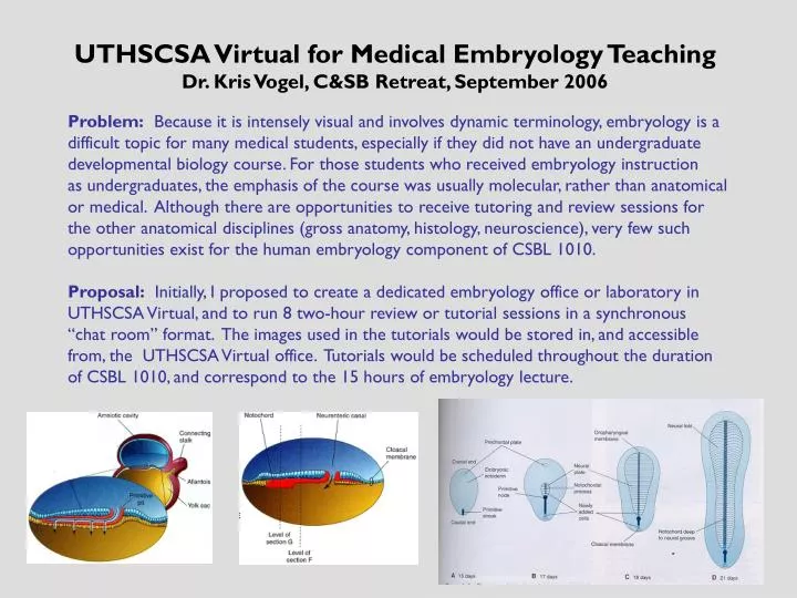
UTHSCSA Virtual for Medical Embryology Teaching Dr. Kris Vogel, C&SB Retreat, September 2006. Problem: Because it is intensely visual and involves dynamic terminology, embryology is a difficult topic for many medical students, especially if they did not have an undergraduate

E N D
UTHSCSA Virtual for Medical Embryology Teaching Dr. Kris Vogel, C&SB Retreat, September 2006 Problem: Because it is intensely visual and involves dynamic terminology, embryology is a difficult topic for many medical students, especially if they did not have an undergraduate developmental biology course. For those students who received embryology instruction as undergraduates, the emphasis of the course was usually molecular, rather than anatomical or medical. Although there are opportunities to receive tutoring and review sessions for the other anatomical disciplines (gross anatomy, histology, neuroscience), very few such opportunities exist for the human embryology component of CSBL 1010. Proposal: Initially, I proposed to create a dedicated embryology office or laboratory in UTHSCSA Virtual, and to run 8 two-hour review or tutorial sessions in a synchronous “chat room” format. The images used in the tutorials would be stored in, and accessible from, the UTHSCSA Virtual office. Tutorials would be scheduled throughout the duration of CSBL 1010, and correspond to the 15 hours of embryology lecture.
Why do we teach embryology to first-year medical students? Carlson (2002) has reviewed many of the reasons for studying embryology as part of the medical curriculum, including understanding the genesis of common birth defects, identifying the origins of the gross anatomical patterns and organization of the body, and understanding advances in reproductive and embryo technologies. How are innovative teaching projects funded? * Innovative Teaching Grants (UTHSCSA) * Instructional Technology Enhancement Grants (UTHSCSA, AT&T) * Outside sources (e.g. American Association of Anatomists) How is the efficacy of new teaching methods evaluated, and what kinds of options are available for sharing and publishing educational research? * Quantitative and qualitative educational research (AIS-ERD UTHSCSA) * ACET website (www.uthscsa.edu/acet/index.asp) * Journals that publish educational research: The Anatomical Record, Academic Medicine, Medical Education, Journal of Dental Education, BMC Medical Education, American Journal of Physiology, etc. t Carlson BM (2002). Embryology in the medical curriculum. Anat. Rec. (New Anat.) 269:89-98.
Methods: What is UTHSCSA Virtual? UTHSCSA Virtual is an online environment designed to facilitate academic collaboration, is accessible from any computer in the world with a current Internet browser, and is available to all UTHSCSA faculty, staff, and students. Some of the options include virtual meetings with transcripts, a virtual reference librarian, an interactive shared desktop, secured document repositories, and a personal office with image repository. * Special thanks to Drs. Aggie Manwell-Jackson and Jim Bower, and to Michael Ottmers (UTHSCSA Virtual)
Screenshot of my UTHSCSA Virtual Embryology Office This is the synchronous, or “chat room” venue. When a student logs in and enters the virtual office, his or her avatar appears in the room. The stage of the microscope is the entry point for the image repository and message board; the chair works as a shared desktop for synchronous discussion.
Screenshots of the asynchronous, or “message board”, venue. I can upload images and text to start threads; students can access these threads at any time, and view or download images. The students can also leave questions or comments as replies in each individual thread. Students can also e-mail questions to me at virtual.embryology@gmail.com Each thread has a descriptive title, and a thumbnail version of the image. So far, I’ve uploaded images corresponding to the first module (5 embryology lectures). Some of the images are simplified or redesigned diagrams from Moore and Persaud, and a few are photos of 3D models I created using Sculpey II (polymer clay). In addition, I’ve used Edward Tufte’s information design principle of “small multiples” to generate summary sheets for visual concepts and terminology.
Methods: “Tufte-izing” embryology diagrams from the Moore and Persaud textbook, i.e. applying principles of information design to medical education “Escaping Flatland”, or design strategies that sharpen the information resolution of paper and the video screen: 1. Increase the number of dimensions that can be represented 2. Increase the data density (amount of information/unit area) Visual displays of information should encourage a diversity of individual viewer styles and rates of editing, personalizing, and understanding. * from Tufte, Edward R. Envisioning Information (Graphics Press, 1990) www.edwardtufte.com
In some cases, the Moore and Persaud Developing Human textbook does an excellent job of information design. These “small multiples” of neurulation in transverse section only required a little tweaking to present a more basic concept, and to make the terminology consistent. Sometimes the textbook organization is not the same as that of the lectures. Therefore, I included a diagram and description of ossification centers in the developing vertebrae, to go along with my lecture on the gross anatomy of the back, vertebral column, and spinal cord.
Direct methods can be used to increase the number of dimensions that can be represented. Here, I’ve constructed polymer clay models of embryos and photographed them, to display 3 dimensions. The bilaminar disc model (above) is one of three models in a series, so that the fourth dimension of time can also be represented. The neurula model on the right was also photographed from the ventral view to display mesoderm and endoderm.
Summary of Second Week of Human Development Design principles used: * Small multiples- similar graphic design allows viewer to focus on changes that occur * Data compression- fit entire second week onto one sheet, so that key points can be compared within eyespan * Color- here, color is used simply to label and identify * Narratives of space and time- emphasis on changes that occur from day to day
Summary of Eye Development * Similar design principles (small multiples, data compression, color) * Self-quiz on congenital eye defects included in text for thread (can be viewed side-by-side with image on computer monitor) * Addressing different learning styles in the text associated with the image: Premature infants may have persistent pupillary membrane, which appears as weblike connective tissue over the pupil. Because of the improvements in caring for very young premature infants (born at 25-26 weeks of gestation), some of your tiniest patients may still have fused eyelids. Once the eyelids begin to open (normally during fetal life), the bulbar and palpebral conjunctivae adopt their proper positions.