Download
1 / 47
470 likes | 601 Vues
Cardiovascular Structure & Function. Cardiovascular system: The heart Arteries Veins Capillaries Lymphatic vessels. Weighting of the heart ceremony: Ancient Egyptians. William Harvey and Blood Flow. April 1, 1578 – June 3, 1657. Introduction.

E N D
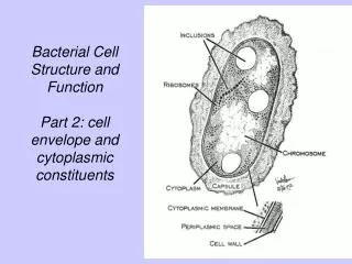
Cardiovascular Structure & Function
Cardiovascular system: • The heart • Arteries • Veins • Capillaries • Lymphatic vessels
William Harvey and Blood Flow April 1, 1578 – June 3, 1657
Introduction • The most basic functions of the cardiovascular system are: • To deliver oxygen and nutrients to body tissues • To remove waste • To regulate temperature
Circulatory system 1. Left ventricle 2. Aorta 3. Arterial system 4. Vena cava 5. Right atrium 6. Right ventricle 7. Pulmonary artery 8. Lungs 9. Pulmonary veins 10. Left atrium 1. Left ventricle
M P A T Heart valves Pulmonary Aortic Mitral Tricuspid
Systolic and Diastolic phases Diastolic phase = Ventricular filling Systolic phase = Ventricular ejection
Cardiac cycle Systole Diastole Animation: http://library.med.utah.edu/kw/pharm/hyper_heart1.html
PA 100 Pressure (mmHg) 50 PLV 0 160 120 LV volume (mL) 80 Ejection Aortic and Ventricular Pressures
Normal pressures Unit = mmHg [1 mmHg = 133 Pa] Left Atrium 2-11 Left ventricle 90-140 / 5-12 Aorta 90-140 / 60-90 Right Atrium 0-8 Right ventricle 15-30 / 0-8 Pulmonary artery 15-30 / 4-12
Hypertension Hypertension is defined as an abnormal increase in arterial pressure Hypertension results in an increase in ventricular work and ventricular hypertrophy
Systolic Pressure 100 Diastolic Pressure mmHg 50 0 0 0.5 1 1.5 400 mL/s 200 0 0 0.5 1 1.5 time in s Pressure and Flow
Classification of arterial pressure (> 18 years) Systolic pressure = maximal pressure Diastolic pressure = minimal pressure CATEGORY SYSTOLIC (mmHg) DIASTOLIC (mmHg) Normal < 130 < 85 Normal ++ 130-139 85-89 Hypertension STAGE 1 (Mild) 140-159 90-99 STAGE 2 (Moderate) 160-179 100-109 STAGE 3 (Severe) 180-209 110-119 STAGE 4 (Very Severe) >209 >119 A very low pressure should also by examined by a Doctor.
3 2 2 4 4 4 3 3 2 1 1 1 1 1 Ventricular pressure-volume curves 120 100 100 Pressure (mmHg) 80 50 Ventricular pressure (mmHg) 60 Stroke volume 0 40 0 0.2 0.4 0.6 0.8 20 160 0 140 60 80 100 120 140 160 Stroke volume 120 Ventricular Volume (mL) Ventricular volume (mL) 100 • Isovolumetric contraction • 2. Ventricular ejection • Isovolumetric relaxation • Ventricular filling 80 0 0.2 0.4 0.6 0.8 Time (s)
120 100 80 Ventricular Pressure (mmHg) 60 40 20 0 60 80 100 120 140 160 Ventricular volume (mL) Ventricular work 1 J P : Ventricular pressure V : Ventricular volume
200 200 Normal pressures 100 100 1 J 0 0 0 0.5 1 1.5 80 120 160 200 200 Severe hypertension 100 100 1.5 J 0 0 0 0.5 1 1.5 80 120 160 Effect of systemic hypertension
SP (systolic pressure) PP MAP (mean pressure) DP (diastolic pressure) Mean pressure (MAP) and pulsed pressure (PP) MAP 2/3 DP + 1/3 SP PP= SP DP
Resistance and compliance Resistance : R MAP / CO MAP : mean arterial pressure CO : cardiac output Compliance : C SV / PP SV : stroke volume PP : pulse pressure Normal conditions (120/80 mmHg) : R = 1.1 mmHg.s/mL C = 2.0 mL/mmHg Severe hypertension (190/115 mmHg) : R = 1.7 mmHg.s/mL C = 0.8 mL/mmHg
Resistance and compliance Normal flow rate : SV = 70 mL, CO = 5L/min Resistance : R MAP / CO R increases MAP increases Compliance : C SV / PP C decreases PP increases MAP 100 PP mmHg 50 0 0 0.5 1 1.5
Hypertension and aortic pressure Normal (120 / 80) 200 R = 1.1 mmHg.s / mL C = 2.0 mL / mmHg MAP = 95 mmHg PP = 40 mmHg R = 1.7 mmHg.s / mL C = 0.8 mL / mmHg MAP = 140 mmHg PP = 75 mmHg MAP 100 95 PP 0 0 0.5 1 1.5 Severe hypertension (190 / 115) 200 MAP 140 PP 100 0 0 0.5 1 1.5
Arterial Pressure Flow, resistance and compliance of the arterial system are significant determinants of arterial pressure Another parameter that can determine arterial pressure is wave reflection in the arterial system.
Arterial pressure and wave reflection aorta Femoral a artery dorsalis pedis
. . . . . . . . . . . . . . . . . . . . . . . . . . . . . . . . . . . . . . . . . . . . . . . . . . . . . . . . . . . . . . . . . . . . . . . . . . . . . . . . . . . . . . . . . . . . . . . . . . . . . . . . . . . . . . . . . . . . . . . . . . . Arterial pressure and wave reflection Incident wave Reflected wave
Heart Sounds Two heart sounds can be distinguished: S1 and S2. S1 is synchronized with early systolic phase. S2 is synchronized with the end of systolic phase. The frequencies are between 10 and 500 Hz with low intensity
1st stethoscope • 1816 Laennec was examining a young female patient • He was embarrassed to place his ear to her chest • Rolled up 24 sheets of paper, placed one end to his ear and the other end to the woman’s chest • Discovered that the sounds were also louder and clearer
Examples of stethoscopes In 1819, the French doctor René Laënnec invented the stethoscope.
Examples of stethoscopes En 1851, Dr. Marsh developed the 1st commercial model.
Pathological heart sounds Several pathologies induce turbulence in the flow in the cardiovascular system. High frequencies in the turbulent flow generate audible sounds.
Aortic stenosis Animation: http://www.healthcentral.com/heart-disease/valves/
Aortic valve Normal vs Stenosed
Congenital Rhumatismal Calcification Aortic stenosis : different origins
Aortic stenosis The most common valvulopathy in industrial countries. Prevalence 1 to 2 % in people > 65 years old. Valvular surgeries in Canada (1996-1997)
Aortic stenosis Carabello et al. NEJM, 1997
Symptoms • Angina pectoris • Syncope • Intolerance to exercise
Investigation of the severity of aortic stenoses • Gorlin Equation • Energy loss coefficient
Biological valves : Xeno-valves : pericardial or porcine Homo-valves : human Ross procedure • Mechanical valves : Cage-ball : Starr-Edwards Mono-leaflet : Björk-Shiley Bi-leaflet : St-Jude Valve replacement
Bioprosthetic valves • Pericardial valves • Porcine valves
Caged-ball : • 1960 (Starr-Edwards) • Mono-leaflet : • Bi-leaflet : Mechanical valves
Stenosed valve Mechanical valve Surgery http://heart-surgeon.com Animation: http://www.healthcentral.com/heart-disease/valves/
Cardiovascular Structure & Function