Download
1 / 30
380 likes | 1.02k Vues
Anatomy of the Sensory Organs. Sensory Function and Vision…. The General Senses…. Sensory receptors specialized cells that monitor the environment and relay information to the CNS. Free nerve endings are the simplest type: they are the dendrites of sensory neurons

E N D
The General Senses… • Sensory receptors • specialized cells that monitor the environment and relay information to the CNS. • Free nerve endings are the simplest type: they are the dendrites of sensory neurons • Complex receptors (eyes) are housed in organs • Some receptors respond to only one kind of stimulus
All sensory receptors send info to the CNS via an action potential… • At the CNS, info is routed according to the stimulus and its location • The stronger the stimulus, the higher the frequency of action potentials • Some receptors adapt, that is their sensitivity to a stimulus is reduced if the stimulus is continually applied (smell) • The RAS can heighten or reduce awareness of sensory information
General versus special senses… General sense receptors included those for temperature, pain, pressure, touch, vibration & proprioception (body position) These receptors are very simple in nature
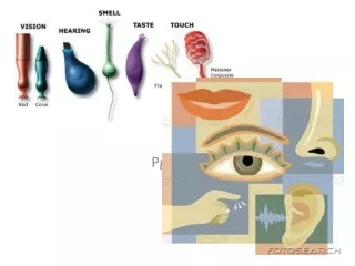
“Special” senses Special senses monitor vision, hearing, olfaction, gustation, and equilibrium through specialized sense organs These sense organs are highly specialized
“General sense” receptors… • Are simple, found everywhere and are classified by their stimulus: • Nociceptors – sensitive to pain • Thermoreceptors – respond to heat • Mechanoreceptors–respond to touch/pressure • Chemoreceptors – response to chemicals • Only 1% of the info they provide ever reaches the cerebral cortex (our consciousness)
Nociceptors/Pain receptors: • found in joints, periosteum of bone and skin • do not adapt! • 2 types of axons carry the painful sensations: • Fast pain sensations (localized, shooting pain) are carried by myelinated axons • Slow pain sensations (generalized, aching pain) are carried by unmyelinated axons: it is difficult to pinpoint the stimulus location • Referred pain is the perception of pain coming from a body area that is NOT stimulated: • Pain originating in viscera is felt on body surfaces
Referred pain: For example, pain produced by a heart attack may feel as if it is coming from the arm because sensory information from the heart and the arm converge on the same nerve cells in the spinal cord.
Thermoreceptors… • Free nerve endings in skin, muscle, liver, and hypothalamus • they adapt quickly • Cold receptors respond to temps <50 • 4X’s as numerous as warm receptors • Warm receptors respond to temps >113 • Both are structurally identical
Mechanoreceptors… • Membrane distortion opens mechanically regulated ion channels to create impulses. There are 3 classes: • Tactile – respond to touch • Baroreceptors – respond to pressure • Proprioceptors – respond to changes in body position
Tactile receptors… • May be simple or complex, superficial or deep, fine (provide detailed information) or crude (provide little information) • Merkel’s – fine touch and pressure • Pacinian – deep pressure • Meissner’s – fine touch and pressure in select areas • Ruffini – pressure or distortion in deep dermal layers
stretch receptors that monitor changes in organ pressure in distensible organs rapidly adapting Generate an AP from their dendrites when organs are stretched or change position They monitor BP, respiration, digestion, and urinary control Baroreceptors are….
Chemoreceptors… Only respond to dissolved chemicals • Rapidly adapting: found in olfaction, taste & the CNS at: • Medulla – receptors are sensitive to pH/CO2 changes in CSF– trigger respiratory adjustments • Aortic/Carotid bodies – sensitive to changes in pH/CO2/O2 blood levels – trigger adjustments in respiration and cardiovascular activity
Proprioceptors…. • Monitor joint position & muscle contraction • DO NOT ADAPT • Structurally complex – are 2 types • Tendon organs – monitor tendon strain • Muscle spindles – monitor muscle length • Most information from these receptors is monitored subconsciously • Are vital for normal skeletal motor function
Olfaction -The Sense of Smell • Consists of paired olfactory organs made of epithelium containing olfactory receptors, supporting cells & stem cells • the receptor cells are highly modified neurons (chemoreceptors) • Contains olfactory glands that produce a multifunctional mucus • Sensory Pathway: olf.epithelium>olf.bulbs>olf.tracts>cortex • http://www.bbc.co.uk/science/humanbody/body/factfiles/smell/smell_ani_f5.swf
Gustation….taste • http://www.bbc.co.uk/science/humanbody/body/factfiles/taste/taste_ani_f5.swf • Taste buds are organs containing gustatory & supporting cells that lie within papillae • Chemicals contact taste hairs which change the MP of taste cells & leads to an AP in the sensory neuron • 4 primary taste sensations – sweet, salt, sour, bitter • Sensory Pathway: sensory receptors>medulla> thalamus>primary sensory cortex
A complex sensory organ: the eye. • is surrounded by accessory structures that act to protect, lubricate, and support it • is a light, compact, durable, and highly specialized hollow organ that weighs about 8 oz and measures 1 inch in diameter. • is divided into anterior (aqueous) & posterior (vitreous) cavities. • its walls are made of 3 “tunics”
Accessory structures of the eye… eyelids (palpebrae) eyelashes & brows exocrine glands lacrimal apparatus Conjunctiva 6 extrinsic occulomotor muscles: the inferior, superior, lateral and medial rectus muscles the superior and inferior oblique muscles
Eye anatomy….. • http://www.macula.org/anatomy/eyeframe.html • The hollow eye is divided into 2 cavities: • An anterior cavity which contains aqueous humor • A posterior cavity which holds vitreous humor • Humors act to stabilize eye shape and provide nutrients
The Tunics of the eye… • Fibrous- the sclera & anterior cornea • Vascular – contains blood vessels, lymphatics, choroid & intrinsic muscles of the iris &ciliary bodies (they support the lens) • Neural – the retina, it contains the rods and cones (photoreceptor cells), bipolar &ganglion cells
Retinal organization … • The retina is made of several cell layers: • Photoreceptor cells – rods lie along the periphery & cones lie at the back of the retina • Bipolar cells synapse with the rods and cones • Ganglion cells synapse with the bipolar cells • The axons of the ganglion cells form the optic nerve • http://www.macula.org/anatomy/retinaframe.htmlhttp://www.macula.org/anatomy/anatomy.html
Macula lutea – area on the retina where the visual image forms, it contains only cones with the greatest numbers at the fovea centralis Optic Disc or “blind spot” is the area where the ganglion cell axons exit the eye to form the optic nerve
Other structures of the eye: • Lens – held in place by suspensory ligaments, it functions to focus the visual image onto the retina • Cornea – clear portion of the fibrous tunic it is contiguous with the sclera • Iris – part of the vascular tunic, it contains blood vessels, pigment, and 2 smooth muscle layers to control the width of the pupil. • Ciliary body – a thick region of the choroid that encircles the lens and supports the suspensory ligments of the lens
Accommodation- focusing an image on the retina by changing lens shape • light bends/refracts as it passes from 1 medium to another. In the eye it goes through the cornea, a. humor, lens, & v. humor. The refraction of light is constant through all but the lens • The lens changes shape to keep the image focused on the retina for greatest visual acquity • Accommodation occurs with response to light and to the distance of the object being viewed • http://www.kscience.co.uk/animations/eye.swf
The Physiology of Vision…How is it that we see? • Photoreceptors respond to visible light: • Rods – sensitive to photons (energy) but not their wavelength (color) allow for vision at night/dim light • The 3 types of Cones (green, red, blue) need bright light & are responsive to wavelength: they allow us to “see” color • Lack of functional cones = colorblindness
The visual pathway…. • Photoreceptor stimulation • Bipolar cell activation • Stimulation of ganglion cell’s whose axons form the… • Optic nerve that cross at the diencephalon and goes to the thalamus that routes info to the visual cortex of occipital lobe and the reflex centers of brain stem • At the optic chiasm, a partial crossover of nerve fibers occurs • http://www.sumanasinc.com/webcontent/anisamples/neurobiology/visualpathways.html