Download
1 / 31
320 likes | 654 Vues
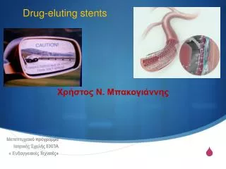
Drug-eluting stents. Χρήστος Ν. Μπακογιάννης Επίκουρος ΚαθηγητήςΑγγειοχειρουργικής Πανεπιστημίου Αθηνών Α΄ Χειρουργική Κλινική ΕΚΠΑ Λαϊκό Νοσοκομείο. Μεταπτυχιακό πρόγραμμα Ιατρικής Σχολής ΕΚΠΑ « Ενδαγγειακές Τεχνικές» 7 /03/1 4. Endothelial injury post implantation. Implanted stent.

E N D
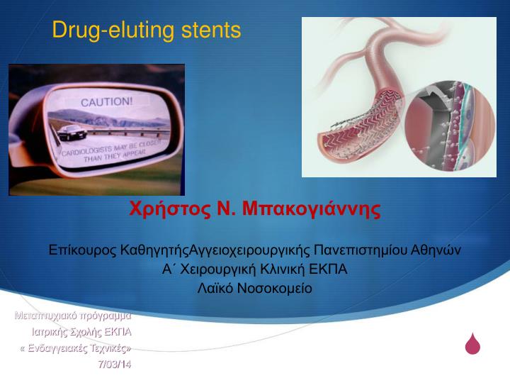
Drug-eluting stents Χρήστος Ν. Μπακογιάννης Επίκουρος ΚαθηγητήςΑγγειοχειρουργικής Πανεπιστημίου Αθηνών Α΄ Χειρουργική Κλινική ΕΚΠΑ Λαϊκό Νοσοκομείο Μεταπτυχιακό πρόγραμμα Ιατρικής Σχολής ΕΚΠΑ « Ενδαγγειακές Τεχνικές» 7/03/14
Endothelial injurypost implantation Implanted stent Plaque Stent implantation causes arterial injury, which can initiate restenosis. The restenosisprocess includes inflammation, migration of smooth muscle cells, smooth muscle cellproliferation and extracellular matrix formation.
Platelet aggregationand activation Drug-eluting stent struts Platelets Red blood cells Inflammatory cells Platelet deposition and activation occur at the injury site, leading to the release ofcell-signaling molecules.
Transmigration ofinflammatory cells Endothelial cells Transmigration ofinflammatory cells Smooth muscle cells Inflammatory cellssecreting cell-signalingmolecules Once activated, these inflammatory cells roll across the endothelial surface andtransmigrate into the lesion.
Activation of smoothmuscle cells Smooth muscle cell extracellular view Cell signaling molecules activatesmooth muscle cells Smooth muscle cellsurface receptor The activated inflammatory cells secrete molecules that bind to specific receptorson smooth muscle cells.
Activation of smoothmuscle cells Smooth muscle cell intracellular view Activatedsmooth musclecell receptor mTOR activatessmooth musclecells to entercell cycle Bound smooth muscle cell receptors activate various intracellular smooth musclecell proteins. One such protein, mTOR, plays a central regulatory role in the cell cycle.
Activation of smoothmuscle cells (III) Cell responds to growth factor stimulation Mitosis Cell resting phase Restriction point DNA synthesis Cell prepares for mitosis Activated mTOR stimulates smooth muscle cells to advance from the G1 phase tothe S phase where DNA replication occurs, causing the smooth muscle cells toundergo mitosis (ie, cell proliferation).
Differential Events Leading to In-Stent Restenosis 1 Fractionof Maximal Response 0 Time Platelet Deposition Leukocyte recruitment VSMC migration / proliferation Matrix deposition
There are three major components to a drug-eluting stent: • Type of stent that carries the drug coating • Method by which the drug is delivered (eluted) by the coating to the arterial wall (polymeric or other) • The drug itself – how does it act in the body to prevent restenosis? • Cordis CYPHER™ sirolimus-eluting stent • Boston Scientific TAXUS™ paclitaxel-eluting stent system, • Medtronic's Endeavor stent which uses ABT-578 • XIENCE PRIME Everolimus Eluting Coronary Stent System
Drug-eluting stents στην SFA Duda SH. Circulation2002; 106:1505–1509.
Τύποι drug-eluting stents με εφαρμογή στην αγγειοχειρουργική
N N N O Chiral N O H H C H 3 O H H O N C H 3 O O O O H O H C 3 O H C 3 H O H H C 3 H C C H H C O 3 3 3 O O H C 3 C H 3 Rapamycin Analogs EVEROLIMUS SIROLIMUS ABT-578 N N N N N N
SMART stents στην SFA The only study which reported local drug delivery in the SFA was the Sirolimus-Coated Cordis Self-Expandable Stent (SIROCCO) trial, in which sirolimus-coated stents were not significantly superior to uncoated stents Duda SH. Circulation2002; 106:1505–1509. Duda SH.J Vasc Interv Radiol 2005; 16:331–338
SMART stents στην SFA Duda SH.J Vasc Interv Radiol 2005; 16:331–338
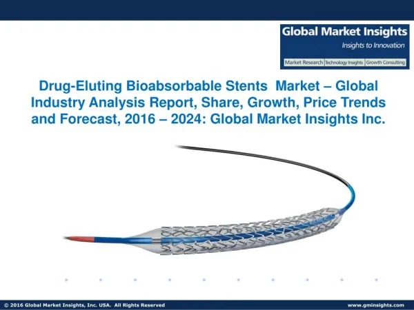
Zilver PTX (paclitaxel) Paclitaxel is a mitotic inhibitor used in cancerchemotherapy. It was discovered in a National Cancer Institute program at the Research Triangle Institute in 1967 when Monroe E. Wall and Mansukh C. Wani isolated it from the bark of the Pacific Yew tree, Taxus brevifolia and named it 'taxol' First, it allows targeted delivery of a drug (paclitaxel) proven to reduce the renarrowing (restenosis) of arteries opened using balloon angioplasty. Second, by eliminating the need for a polymer, Zilver PTX avoids the potential patient risks posed by leaving a permanent foreign, plastic substance in the body. Zilver PTX mechanisms of action: Hydrophobic—PTX won't wash off. It adheres to the stent without the need for a synthetic polymer Lipophilic—PTX seeks the lipids in the vessel wall and attaches Antiproliferative—once in the cell, PTX blocks cell division (proliferation) for the life of the cell
Διαφορετική αποτελεσματικότητα drug-eluting stents στην SFA & στα στεφανιαία.ΓΙΑΤΙ; the distance between the stent struts of the Smart stent was much larger compared to the Cypher stent, leading to a lower drug dose in the SFA compared to the coronary arteries Oliva VL. J Vasc Interv Radiol. 2005;16:313–315.
Drug-coated balloons for femoropopliteal PTA: Paccocath (Cotavance) balloon) Scheller B et al. Circulation. 2004;110:810–814. Scheller B et al. N Engl J Med. 2006;355:2113–2124. Scheller B. EuroIntervention. 2008;4(suppl C):C63–C66. Scheller B et al. Heart. 2007;93:539–541.
Local Taxane with Short Exposure for Reduction of Restenosis in Distal Arteries (THUNDER) trial • 154 patients (24% smokers, 49% diabetics) with femoropopliteal lesions • Paccocath (n=48 patients) • no adverse event • 6 months mean late lumen loss 0.461.2 mm vs. 1.761.8 mm for controls (p=0.001) • 6-month & 12-month angiographic binary restenosis were 10% and 25% for the Paccocath patients vs. 41% and 59% for the control patients (p=0.01) Currently, the use of antiproliferative agents, either exposed by stents or balloon catheters in preventing restenosis in infrainguinal arteries, is still investigational. Tepe G, et al. N Engl J Med.2008;358:689–99.
Ανεπιθύμητες ενέργειες • Vascular toxicity rather than cytotoxicity • Late incomplete apposition • Medial thinning • Aneurysm/rupture • Delayed re-endothelialization Vasculo-toxic effects in pig coronaries: 90 days High dose, fast release Low dose, slow release Rogers C et al. Circ. 2000.
Late incomplete apposition Potential for stent thrombosis Follow-up Baseline In a Taxus and Cypher study of patients with late incomplete apposition upon clopidogrel discontinuation: 20% had stent thrombosis* No remodeling Positive remodeling
Taxus and Cypher BMS 1 2 3 4 5 6 7 8 9 11 15 16 17 20 > 40 • Conclusions: • DES (solid line) consistently show less endothelialization compared with BMS (dashed line) regardless of time point, even beyond 40 months • DES are not fully endothelialized, whereas BMS are completely covered by 6 to 7 months Percent struts endothelialized Human analysis: DES vs BMS 100 90 80 70 60 50 Percentage endothelialization 40 30 20 10 0 Duration in months Joner, Virmani et al. Circulation. 2005;112:3210.
Exposed stent struts at 6 months > 80% Cypher struts exposed vs BMS struts 100 Sirolimus-eluting stent 75 50 Percent 25 0 Incomplete coverage Complete coverage Grade 0 Grade 1 Grade 2 Grade 3 0 25 50 Percent 75 Bare-metal stent 100 Kotani et al. JACC. 2006;47:2108-2111.
Endothelial dysfunction Reduction in eNOS and nitric oxide (NO) production • Normal vessels dilate in response to exerciseor acetylcholine (ACH) This response is dependent on endothelial production of NO • Atherosclerotic vessels are characterized byhaving endothelial dysfunction and constrictin response to exercise or ACH Cai H, Harrison DG. Circ Res. 2000;8This is explained by either a loss ofendothelial cells or loss of eNOS expressionand NO production 7:840-844. Bonetti PO et al. ATVB. 2003;23:168-175.
Σασευχαριστω για την Προσοχησασ! Μεταπτυχιακό πρόγραμμα Ιατρικής Σχολής ΕΚΠΑ «Ενδαγγειακές Τεχνικές» 7/03/14