Download
1 / 40
460 likes | 546 Vues
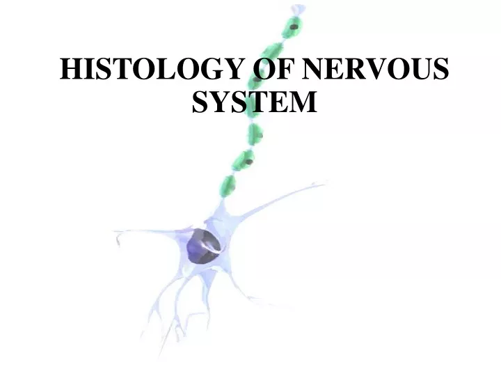
HISTOLOGY OF NERVOUS SYSTEM. N ervous S ystem. The most complex system in the human body . Formed by network more than 100 million neuron . Each neuron has a thousand interconnection a very complex system for communication .

E N D
Nervous System The most complex system in the human body. Formed by network more than 100 million neuron. Each neuron has a thousand interconnection a very complex system for communication. Nerve tissue is distribute throughout the body, anatomically divide into : CNS & PNS. Structurally consist : nerve cells & glial cells
Cells of Nervous System NEURON NEUROGLIA
Structure of Neuron • Principle cells of Nervous Tissue. • Consist of 3 parts : • CELL BODY (perikaryon/soma) • A single AXON • Multiple DENDRITES • 5-150 µm
Cell Body (Perikaryon) • Central portion of the cell. • Generally are polygonal. • Different shape and size characteristic regions of nervous system. • Contain : • Nucleus • Perinuclear cytoplasm
Ultrastructure of Neuron Nucleus : • large, spherical to ovoid and centraly located. • a single prominent nucleolus. • Finely dispersed chromatin.
Ultrastructure of Neuron Cytoplasm: • Abundantof R.E.R • Polyribosomes • Basic dyes (a+b) Nissl Bodies • lots of S.E.R. • Golgi bodies (perikaryon) • protein secreting cell
Ultrastructure of Neuron • Many mitochondria, most abundant in axon terminal • Extensive cytoskeleton axonal transport • One centriole do not undergo cell divisions
Dendrite and Axon • Axon: • Single process up to 100 cm • Originate from axon hillock • Devoid ribosome • Dilatation of distal portion axon terminal end bulbs synapse • conducting impulse away from the soma • Axonal transport • Dendrites: • Multiple elongated processes • Cytoplasmic~perikaryon (devoid golgi complex) • Receiving stimuli
Neurons Classification According to the size and shape of the processes: • Multipolar: the most abundant, Ex: pyramidal cells, Purkinje cells. • Bipolar: Ex: visual, auditory system. • Pseudounipolar: Ex: sensoryganglia.
Neurons Classification According to the size and shape of the processes
Neurons Classification According to their function: • Sensory Neuron (afferent): • Receive sensory input conduct impulses to CNS • Motor Neuron (Efferent): • CNS conduct impulses to muscles, glands and other neurons • Interneuron: • In the CNS as interconnectors, establish neuronal circuit between sensory and motor neuron
Neuron Grouping • CORTEX: • Neuron form six layers on the cerebrum. • Form three layers on the cerebellum. • NUCLEI: • In subcortical region (thalamus, midbrain, brainstem and spinal cord) neuron form irregular cluster nuclei • GANGLION: • Cluster of neuron outside the CNS
Synapses • Sites of impulse transmission. • Convert electrical signal into chemical signal • Permit neurons to communicate. • Types of synapses : • Axodentritic synapse. • Axosomatic synapse. • Axoaxonic synapse. • Dendrodentritic synapse.
Neuroglial Cells • Metabolic and mechanical supportfor neuron. • 10 times abundant than neurons. • Neuroglial cells undergo mitosis. • Function: provide neurons with structural support and maintain local conditions for neuronal function. • Staining: silver or gold impregnation, histochemical technique. • Classification: • Oligodendrocytes • Astrocytes • Ependymal Cells • Microglia • Schwan cells CNS PNS
Neuroglial Cells • Astrocytes: • Pedicles binds to capillaries and to the pia mater form glial limitans. • Controlling the ionic & chemical environment of neurons • Energy metabolism • Form cellular scar tissue • Form the blood-brain barrier
Neuroglial Cells • Protoplasmic astrocytes: • Granular cytoplasm. • Envelop the surface of nerve cells and blood vessels. • Fibrous astrocytes: • Long processes. • Predominantly in white matter.
Neuroglial Cells • Oligodendrocytes: • Produce myelin sheath. (electrical insulation) in CNS. • A single cell wrap several axons (40 to 50). • Form nodes of Ranvier
Neuroglial Cells • Microglia: • Phagocytic cells, scattered throughout the CNS. • Derived from mesoderm. • Small cell bodies. • Their nuclei have elongated shape. • Short processes with small expansions –thorny appearance. • Functions: Clearing debris, Act as APC, protect the CNS from viruses and microorganism.
Neuroglial Cells • Ependymal Cells: • Low columnar ciliated epithelial cells line the ventricles of the brain & central canal spinal cord. • Formation of choroid flexus produce CSF. • Facilitates the movement of CSF.
Neuroglial Cells • Schwann cells: • Analogue to Oligodendrocyte. • Produce myelin sheath in the PNS.
Nervous System is anatomically divided in to: • Central nervous system (CNS). • Peripheral nervous system (PNS).
The CNS • Consist of : • Cerebrum • Cerebellum • Spinal cord • No connective tissue soft, gel like • When sectioned : • White matter • Gray matter • Covered by meninges
Cerebrum • Gray Matter: • Contains neuronal cell bodies, dendrites and glial cells • Six layers composed of neuron • White Matter: • Contains myelinated axons and myelin-producing oligodendrocytes
Cerebellum • Gray Matter: • Three layers: • Outer molecular layer • Central layer of large Purkinje cells • Inner granule layer • White Matter: • The same as cerebrum
Spinal Cord • Gray Matter (central) shape of “H” • Central canal lined by Ependymal cells • Legs of the “H” form : • Anterior horns • Posterior horns • Neurons : large and multipolar • White Matter (peripheral)
The PNS • Bundles of nerve fibers (axons) outside the CNS & surrounded by connective tissue. • Main component: • Peripheral nerves • Ganglia • Nerve endings
Nerve Fibers • Consist of axons enveloped by a special sheath. • Group of fibers constitute the peripheral nerve. • Two types: • Myelinated fiber • Unmyelinated fiber
Nerve Fibers • Myelinated fibers: • A single Schwann cell wraps around single axon form myelin sheath nodes of Ranvier. • Unmyelinated fibers: • A single Schwann cell envelopes several axon. • Fibers enveloped within simple clefts of Schwann cells
Conduction Velocity Depend on the extent of Myelination: • Unmyelinated fibers • No nodes of Ranvier continuous conduction. • Slower conduction • Myelinated fibers: • Gap of myelin sheath (nodes of Ranvier ) saltatory conduction. • Faster conduction.
Connective Tissue Investments • Epineureum: • Dense collagenous Con. Tissue with thick elastic fiber • Prevent damage by overstreching • Perineureum: • Dense con. Tissue • Isolates neural environment (blood-nerve barrier) • Endoneureum: • Loose con. Tissue • Regulation of microenvironment of nerve fiber
Ganglia • Ovoid structure containing neuronal cell bodies, glial cells supported by connective tissue. • Function : Relay stations to transmit impulses. • Types: • Sensory ganglia • Autonomic ganglia
Ganglia • Sensory Ganglia (cell bodies of sensory neuron) • Unipolar cell bodies enveloped by cuboidal capsule cells • Cranial ganglia: Associated with the cranial nerve • Spinal ganglia: Associated with the spinal nerve • Autonomic Ganglia (cell bodies of postganglionic autonomic nerves) • Multipolar neuron enveloped by satellite cells. • Some are located within certain organ (intramural).
Autonomic Nervous System Sympathetic System Parasympathetic System The nuclei located in the medulla and midbrain and in the sacral portion of spinal cord. Pre ganglionic fibers leave the CNS trough cranial nerve III, VII, IX and X and also trough II, III, IV sacral nerve The ganglion located near the effector organs. The chemical mediator pre and postganglionic fibers is acethilcholine. • The nuclei located in the thoracic and lumbar segment of spinal cord. • Preganglionic fibers leave the CNS by way of ventral roots. • The chemical mediator postganglionic fibers is norepinephrine.
Regeneration of CNS?? • Regeneration nerve fibers in CNS is not possible, because: • An endoneureum is not present • Oligodendrocytes do not proliferate • Astrocytes deposit scar tissue (plaque)