Download
1 / 20
210 likes | 413 Vues
Alzheimer’s Disease. Raquel Peralta. Introduction. Alzheimer’s disease (AD) is the most common form of dementia An estimated 4 million Americans suffer from AD. The disease usually begins after age 60, and risk goes up with age. In 1907 Dr. Alois Alzheimer, a German doctor identified AD.

E N D
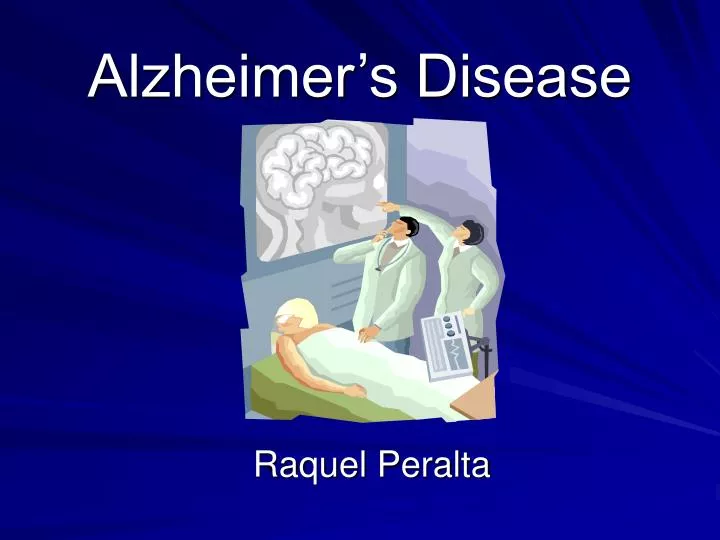
Alzheimer’s Disease Raquel Peralta
Introduction • Alzheimer’s disease (AD) is the most common form of dementia • An estimated 4 million Americans suffer from AD. The disease usually begins after age 60, and risk goes up with age. • In 1907 Dr. Alois Alzheimer, a German doctor identified AD
Hallmarks of AD Example of senile plaques (brown) and neurofibrillary tangles (black) in a brain with AD.
Neurofibrillary tangles (NFT) Tangled tau proteins wound-up in a helix, and located inside of neurons Tau binds to tubulin to form microtubules Microtubules provide support, shape, and nutrient routes within cells Therefore, when the tau proteins become hyperphosphorylated, it causes microtubules to collapse Senile or amyloid plaques Intracellular accumulations of β-amyloid Form hard insoluble plaques Hallmarks cont.
Brain Atrophy AD patient Loss of neurons, and normally convoluted surface of the brain Normal individual of same age
Etiopathogenesis • Currently unknown • There probably is not one single cause, but several factors that affect each person differently • Possible risk factors • Age • Genetics - mutation in chromosome 21 may lead to Familial AD • Apolipoprotein E (apoE) • Diet, environment, and viruses
Symptoms • AD begins slowly • Mild forgetfulness • In the later stages of AD • people may forget how to do simple tasks, like brushing their teeth or combing their hair • problems with speaking, understanding, reading, or writing • Eventually, patients need total care.
Treatments • No treatment can stop AD • For people in the early and middle stages of the disease, the drugs tacrine (Cognex), donepezil (Aricept), may help prevent some symptoms from becoming worse for a limited time • Some medicines may help control behavioral symptoms such as sleeplessness, and agitation • Scientists are testing different types of nonsteroidal anti-inflammatory drugs (NSAIDs), and vitamines
Paper Analysis Sequence of neurofibrillary changes in aging and Alzheimer’s disease: A confocal study with phospho-tau antibody, AD2. Galvan et.al., Journal of Alzheimer’s Disease
Brodmann Areas Area of Focus
1. The hippocampus-important for learning and short-term memory.. 2. Entorhinal cortex - Scientists believe that Alzheimer's dementia begins here.
Purpose • In the present study, neurons of the entorhinal cortex, hippocampus and frontal lobe from non-demented and AD cases were stained in order to study neurofibrillary changes.
Immunolabeling • AD2 • phosphorylation dependent monoclonal antibody to tau • recognizes phosphorylated Ser396 and Ser404 in NFTs • Thiazin red (TR) • histochemical dye • shows binding sites for β-amyloid and tau when in fibrillar states
Materials & Methods • Brains obtained at autopsy (postmortem 7-48 h) • Tissue fixed in 10% formalin solution • Immunocytochemistry reactions – for visualization of immunoreactive products • Confocal microscopy- from each area 5-15 serial Z-sections were collected using the dual channel image system • The density of NFTs was determined by counting 3 different fields randomly chosen in each region.
10 Demented (AD) Ages 69-92 Cognitive status evaluated with the Clinical Dementia Rating (CDR) score. 1 person had mild memory loss 3 moderate 6 severe dementia 10 non-demented Ages 63-93 Cognitive status evaluated with the Clinical Dementia Rating (CDR) score. 6 persons had no memory loss 4 questionable Brains used AD brains were diagnosed according to NINCDS-ADRDA criteria, and neuropathologicaly evaluated according to CERAD criteria. NINCDS-ADRDA=The National Institute of Neurological and Communicative Disorders and Stroke -Alzheimer’s Disease and Related Disorders Association. CERAD=CONSORTIUM TO ESTABLISH A REGISTRY FOR ALZHEIMER'S DISEASE
Double labeling with mAb AD2 and TR in AD brains (fig.1) Patchy perinuclear mAB AD2 stained Granuals stained nucleus Intracellular NFT
Double labeling with AD2 and TR (fig. 2) Intracellular NFT Granulated NFT Senile plaque with B-amyloid deposits Dystrophic neurites
Regional distribution of immunoreactive NFTs in demented and non-demented groups
Schematic showing the stages of mAb AD2 immunoreactivity in tangles
Conclusions • The density of intracellular-NFTs correlates significantly with cognitive impairment. • However, this does not exclude other possible mechanisms (other than NFT formation) involved in neuronal death • The results provide evidence for the sequence of tangle formation as detected by mAB AD2. • NFT occurs in normal aged people, but in lower amounts than in AD cases.