Download

1 / 42
520 likes | 835 Vues
Myopathies. Pathology. Objectives:. At the end of this lecture, the students should be able to: Understand the structure of the various types of muscle fibers. Acquire a basic knowledge of the classification of myopathies and give examples of these disorders.

E N D
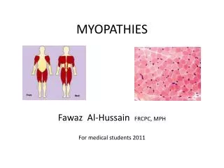
Myopathies Pathology
Objectives: At the end of this lecture, the students should be able to: • Understand the structure of the various types of muscle fibers. • Acquire a basic knowledge of the classification of myopathies and give examples of these disorders. • Understand the meaning of the term muscular dystrophy and have a basic knowledge of the incidence and clinicopathological manifestations of Duchenne's and Becker's muscular dystrophies. • Know the pattern of inheritance of myotonic dystrophy and its clinicopathological presentations.
Contents: • The definition of motor unit and muscle fiber types. • Classification of myopathies. • Muscle atrophy, pathological features and causes. • Neurogenicmyopathy: definition, causes and pattern of nerve injury. • Duchenne and Becker Muscular Dystrophy: incidence, Clinicopathological characteristics, with special emphasis on the rule of dystrophin protein. • Myotonic Dystrophy: definition and main Clinicopathological features with special emphasis of inheritance pattern.
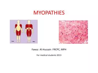
Skeletal muscle Fiber types • Depending on the nature of the nerve fiber doing the enervation, the associated skeletal muscle develops into one of two major subpopulations • A single "type I" or "type II" neuron will innervate multiple muscle fibers and these fibers are usually randomly scattered in a "checkerboard pattern" within a circumscribed area within the larger muscle
Skeletal muscle Fiber types • The different fibers can be identified using specific staining techniques: • type I: • "slow twitch“ • more dependent on fat catabolism for energy through mitochondrial oxidative phosphorylation • red, refers to this being the dark (red) meat on birds where fiber type grouping in different muscles (e.g., thigh vs. breast meat) is quite pronounced • type II: • "fast twitch“ • more dependent on glycogencatabolism for energy through glycolysis • white
MYOPATHY • Myopathy as a term may encompasses a heterogeneous group of disorders, both morphologically and clinically • Recognition of these disorders is important for genetic counseling or appropriate treatment of acquired disease
Myopathies • Diseases that affect skeletal muscle can involve any portion of the motor unit: • primary disorders of the motor neuron or axon • abnormalities of the neuromuscular junction • a wide variety of disorders primarily affecting the skeletal muscle itself (myopathies)
Myopathies • skeletal muscle disease can be divided into: • Neurogenic • Muscular dystrophies • Congenital • inherited mutations of ion channels • inborn errors of metabolism (e.g. glycogen and lipid storage diseases) • mitochondrial abnormalities • Toxic • Thyrotoxicmyopathy • Ethanol myopathy • Drugs (e.gChloroquine) • Infectious • Disorders of the neuromuscular junction (e.g. myasthenia gravis)
MUSCLE ATROPHY • A non-specific response • Characterized by abnormally small myofibers • The type of fibers affected by the atrophy, their distribution in the muscle, and their specific morphology help identify the etiology of the atrophic changes
MUSCLE ATROPHY • Causes: • Simple disuse, type II fibers • Exogenous glucocorticoids or endogenous hypercortisolism (proximal weakness), type II fibers • Myopathies • Neurogenic atrophy
MUSCLE ATROPHYNeurogenic Atrophy • Neurogenic Atrophy : • Both fiber types • Clustering of myofibers into small groups • Deprived of their normal enervation, skeletal fibers undergo progressive atrophy
MUSCLE ATROPHYNeurogenic Atrophy • Loss of a single neuron will affect all muscle fibers in a motor unit, so that the atrophy tends to be scattered over the field • With re-enervation, adjacent intact neurons engage the neuromuscular junction of the previously de-enervated fibers new connection is established these fibers assume the type of the innervating neuron whole groups of fibers can eventually fall under the influence of the same neuron, and become the same fiber type (fiber type grouping)
MUSCLE ATROPHYNeurogenic Atrophy • In that setting, if the relevant enervating neuron now becomes injured, rather large coalescent groups of fibers are cut off from the trophic stimulation and wither away (grouped atrophy), a hallmark of recurrent neurogenic atrophy
MUSCULAR DYSTROPHY • A heterogeneous group of inherited disorders • Often presenting in childhood • Characterized by progressive degeneration of muscle fibers leading to muscle weakness and wasting • Histologically, in advanced cases muscle fibers are replaced by fibrofatty tissue • This distinguishes dystrophies from myopathies, which also present with muscle weakness
Dystrophin • Dystrophin is a large protein (427 kD) that is expressed in a wide variety of tissues, including muscles of all types, brain, and peripheral nerves • Dystrophin attaches portions of the sarcomere to the cell membrane, maintaining the structural and functional integrity of skeletal and cardiac myocytes • The dystrophin gene (Xp21) spans (∼1% of the total X chromosome), making it one of the largest in the human genome; its enormous size is a probable explanation for its particular vulnerability to mutation
Duchenne and Becker Muscular Dystrophy • X-Linked Muscular Dystrophy • The two most common forms of muscular dystrophy • DMD is the most severe and the most common form of muscular dystrophy, with an incidence of about 1 per 3500 live male births • DMD becomes clinically evident by age of 5, progressive weakness leading to wheelchair dependence by age 10 to 12 years death by the early 20s • Although the same gene is involved in both BMD and DMD, BMD is less common and much less severe
Duchenne and Becker Muscular Dystrophy • Morphology: • The histologic features of DMD and BMD are similar • Marked variation in muscle fiber size (atrophy and hypertrophy) • Range of degenerative changes (fiber necrosis) • Regeneration, including sarcoplasmicbasophilia, nuclear enlargement, and nucleolar prominence • Connective tissue is increased • Abnormal staining for dystrophin • Extensive fiber loss and adipose tissue infiltration
Dystrophin • Deletions appear to represent a large proportion of the genetic abnormalities, with frame shift and point mutations accounting for the rest • Approximately two-thirds of the cases are familial, with the remainder representing new mutations • In affected families, females are carriers; they are clinically asymptomatic but often have elevated serum creatinekinase and can show mild histologic abnormalities on muscle biopsy
Pathogenesis • DMD and BMD are caused by abnormalities in the dystrophin gene • The role of dystrophin in transferring the force of contraction to connective tissue has been proposed as the basis for the myocyte degeneration that occurs with dystrophin defects, or with changes in other proteins that interact with dystrophin
Clinical Features • Boys with DMD: • Normal at birth, and early motor milestones are met on time • Walking is often delayed • Weakness begins in the pelvic girdle muscles and then extends to the shoulder girdle • Enlargement of the calf muscles associated with weakness, a phenomenon termed pseudohypertrophy, is an important clinical finding • The increased muscle bulk is caused initially by an increase in the size of the muscle fibers and then, as the muscle atrophies, by an increase in fat and connective tissue • Pathologic changes are also found in the heart, and patients may develop heart failure or arrhythmias
Clinical Features • Cognitive impairment seems to be a component of the disease and is severe enough in some patients to be considered mental retardation • Serum creatine kinase is elevated during the first decade of life but returns to normal in the later stages of the disease, as muscle mass decreases • Death results from respiratory insufficiency, pulmonary infection, and cardiac decompensation
BMD • Boys with BMD develop symptoms at a later age than those with DMD. The onset occurs in later childhood or in adolescence, and it is accompanied by a generally slower and more variable rate of progression • Although cardiac disease is frequently seen in these patients, many have a nearly normal life span
Mitochondrial myopathies • Can involve mutations in either mitochondrial or nuclear DNA that encodes mitochondrial constituents • Mitochondrial myopathies typically present: • in young adulthood • with proximal muscle weakness • sometimes with severe involvement of the ocular musculature (external ophthalmoplegia)
Inflammatory Myopathies • Inflammatory myopathies make up a heterogeneous group of rare disorders characterized by immune-mediated muscle injury and inflammation • Based on the clinical, morphologic, and immunologic features, three disorders: • Polymyositis • Dermatomyositis • Inclusion body myositis
Homework • Define Myotonia? • What is the inheritance and the mutation pattern that characterize myotonic dystrophy? • What is the clinical presentation of myotonic dystrophy? • Source: Robbins basic pathology, 9th edition