Download
1 / 12
120 likes | 340 Vues
Mathematical Description of the Ocular Response Analyzer Applanation Signal in Post-LASIK Ectasia. Mujtaba A. Qazi, M.D. 1,2 David A. Luce, Ph.D. 3 Jay S. Pepose, M.D., Ph.D. 1,2 1. Washington University School of Medicine 2. Pepose Vision Institute, St. Louis, Missouri

E N D
Mathematical Description of the Ocular Response Analyzer Applanation Signal in Post-LASIK Ectasia Mujtaba A. Qazi, M.D.1,2 David A. Luce, Ph.D.3 Jay S. Pepose, M.D., Ph.D.1,2 1. Washington University School of Medicine 2. Pepose Vision Institute, St. Louis, Missouri 3. Reichert Inc.
Financial Disclosure • Dr. Pepose has received research support from Reichert. • Dr. Luce is an employee with Reichert.
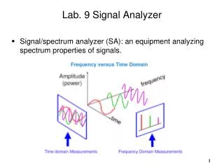
Ocular Response Analyzer • Uses air pulse to produce 2 (inward and outward) applanation events: • AIR PULSE & INFRARED Signals • IOP-g (Goldmann equivalent) • IOP-cc (Cornea Compensation): IOP calculated as a function of CH • Corneal Hysteresis (CH): ability of cornea to dampen or absorb applied force = P1-P2 (mean value in normal population ~ 10) • Corneal Resistance Factor (CRF): ability of cornea to resist or rebound from applied force = P1-0.43xP2 (mean value in normal population ~10)
Study Design • ORA waveforms captured in series of eyes with clinical diagnosis of Post-LASIK Ectasia (n=14) • Compared to pachymetry-matched: • Pre-LASIK, Control eyes from pool of preoperative keratorefractive surgery patients (n=7) • Post-LASIK, Control eyes (n=17) • Keratoconus eyes (n=22) • Along with IOP & Biomechanical metrics, • 38 novel ORA signal parameters evaluated • ORA signals then entered into 11 x 36 Matrix Neural Net
ORA Measurements Pachymetry-matched KCN, pre- and post-LASIK Controls, and post-LASIK Ectasia CRF: Pre-LASIK to LASIK Ectasia approaches statistical significance (p=0.06, ANOVA with Bonferroni correction)
Applanation Waveform analyzed using 38 mathematical metrics, such as: • Pre-LASIK Control • Post-LASIK Control • Post-LASIK Ectasia • Height of Peak 1 & 2 • Width of Peak 1 & 2 • Ratio of Height : Width of Peak 1 & 2 • Slope of the rising and descending portions of Peak 1 & 2 • Acceleration of the slopes of Peak 1 & 2 • Slope of upper half of Peak 1 & 2 • Ascending segments minus descending segments of rising portion of Peak1 & 2 (“chatter”) • Ratio of width of upper half : width of lower half of Peak 1 & 2
Neural Net Analysis There was a statistically significant difference in the mean Neural Net Scores of these 4 populations (p<0.01)
Unoperated Normal Post-LASIK Normal KCN Post-LASIK Ectasia Neural Net Scores
Summary: • There is a trend for lower CRF (and CH) values in the Post-LASIK Ectasia group compared to pachymetry-matched control and Keratoconic eyes. • There is no difference in mean IOPcc for all groups evaluated. • The applanation signals are altered by corneal pathology or surgery. This may be due to a smaller area of applanation or a dynamic misalignment due to non-uniform deformation of the asymmetric corneal surface with air-pulse application. • Novel ORA signal metrics, applied through a Neural Net, may be used to assist in the diagnosis of corneal ectasia. • A larger sample size is needed to fully evaluate the utility of this technique for differentiating between early or forme fruste ectasia and normal eyes and to validate the Neural Net.